Fig. 12.1
MRI of a patient with myocardial thinning on cine and fibrosis on delayed gadolinium-enhanced imaging (DE-MRI) following MI. (a) Cine MRI with 4-chamber view shows myocardial thinning in apical and lateral walls with dyskinesia (arrows) on systole. (b) DE-MRI reveals hyperenhancement (arrows) with transmural involvement in apex and partial thickness involvement in lateral wall
12.3 Specific Imaging Finding for Chronic Ischemic Heart Disease
12.3.1 Left Ventricular Aneurysm
Left ventricular aneurysm is most commonly the result of myocardial infarction, usually involving LV anterior wall.
Hypertrophic cardiomyopathy and Chagas disease can be also causes of left ventricular aneurysm. The aneurysm may be asymptomatic or present as heart failure, sustained ventricular tachyarrhythmias, or arterial embolism (Fig. 12.2) [3].
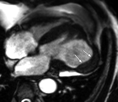
Fig. 12.2
MRI of a patient with pseudoaneurysm of LV in lateral wall. Cine MRI with 4-chamber view shows a large wide-neck aneurysm in lateral wall with thrombus (arrows)
12.3.2 Left Ventricular Pseudoaneurysm
Left ventricular pseudoaneurysm develops after an acute MI which is complicated by a ventricular free wall rupture that is contained by localized pericardial adhesions and generally occurs after inferior myocardial infarction due to occlusion of the left circumflex artery inferior wall (Table 12.1).
True aneurysm
Pseudoaneurysm
Consists of an endocardium, myocardium, and epicardium ± thrombus
Consists of an epi-/pericardium ± thrombus
Wide neck/base
Narrow neck/base
Low risk of rupture
Higher risk of rupture
Commonly anterior wall
Commonly inferior wall
Marked enhancement of the pericardium
12.3.3 Left Ventricular Thrombus
The detection of thrombi after a recent MI is an indication for long-term anticoagulation.
Both MRI and CT imaging can excellently detect thrombus within LV cavity, and MRI has been shown to detect small mural thrombus within apical aneurysm better than contrast echocardiography (Fig. 12.3).
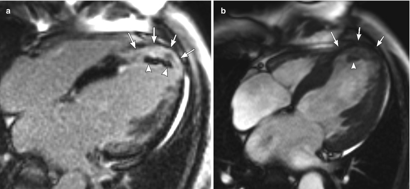
Fig. 12.3
MRI of a patient with myocardial thinning and thrombus. (a) DE-MRI with 4-chamber view shows wall thinning (arrows) with hyperenhancement (arrows) and mural thrombus (arrowheads). (b) Cine MRI with 4-chamber view shows wall thinning (arrows) and thrombus (arrowhead)< div class='tao-gold-member'>Only gold members can continue reading. Log In or Register to continue
Stay updated, free articles. Join our Telegram channel

Full access? Get Clinical Tree


