Fig. 6.1
ECG on presentation at the local hospital two and a half years after surgery showing T-wave inversion in mid-precordial leads
Three months later he developed sudden shortness of breath while walking home; on arrival he collapsed and was coughing blood-stained sputum. He was brought to Accident and Emergency by ambulance, receiving 15 L O2/min by face mask but remained hypoxic, although conscious. Arterial lactate was 4 mmol/l, troponin was grossly elevated and echocardiogram showed globally poor left ventricular function. Chest CT showed no evidence of pulmonary embolus but revealed bilateral lower lobe consolidation. Dobutamine was commenced and urgent cardiac catheterisation arranged. The working diagnosis was myocarditis or coronary embolism, possibly related to poor INR control.
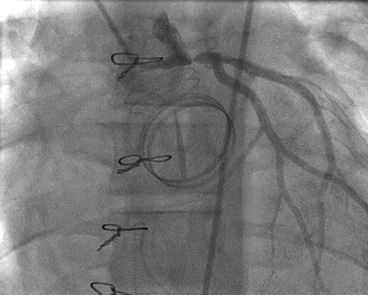
Catheterisation was performed under local anaesthesia and he experienced chest pain during the procedure. The right coronary artery was normal; however, the left main stem was severely narrowed where it crossed over the ring of the On-X valve in the aortic position (Fig. 6.2), and the valves were functioning normally. The following day he was taken to the operating theatre, where he underwent patch repair of the left main stem and grafting of the left internal mammary to LAD. He made an uneventful recovery and left ventricular function gradually improved.


Fig. 6.2
Snapshot of the cardiac catheterization showing mechanical compression of the left main stem at the point of crossing over the ring of the On-X valve in the aortic position
Discussion
Chest pain is common in children and accounts for 10–15 % of ambulatory cardiology visits but in contrast to adult practice very rarely represents cardiac pathology and is usually musculoskeletal, pulmonary, gastrointestinal or pyschogenic in origin (Table 6.1). Cardiac causes of chest pain in children and adolescents are found in less than 5 % of presentations but one must remain alert to the possibility.
Table 6.1
Causes of non-cardiac chest pain in paediatric patients
Aetiology | |
|---|---|
Musculoskeletal (50–68 %) | Costochondritis Muscle strain Trauma Slipping rib syndrome |
Respiratory (3–12 %) | Asthma Pneumonia Bronchitis Pleuritic pain Pulmonary embolus Pneumothorax |
Psychogenic (10–20 %) | Conversion disorder Panic/anxiety attack |
Gastrointestinal (2–8 %) | Oesophagitis Gastritis Gastroesophageal reflux Pancreatitis Gastric ulcer Biliary colic/disease |
Other (<10 %) | Acute chest syndrome of sickle cell disease Skin infection Breast disease |
The lack of evidence-based standards for the evaluation of chest pain in paediatric patients has led to a widespread variation in practice among cardiologists, with exercise stress testing, echocardiography and ambulatory ECG commonly requested. In the absence of any cardiac history all appear to have a very low yield. Previous studies have shown that exercise stress testing was not able to detect any cardiac disorders in otherwise healthy children and adolescents. Ambulatory ECG is also low-yield, except in the presence of palpitations or syncope, and echocardiography is more likely to reveal incidental findings. However, in the presence of a concerning history such as previous cardiac surgery, exertional pain and/or collapse, together with clinical and ECG findings, echocardiography can lead to the diagnosis of significant cardiac lesions such as anomalous coronary artery from the pulmonary artery, cardiomyopathy, pulmonary hypertension, myocarditis, pericarditis, and left severe ventricular outflow tract obstruction. Cardiac biomarkers, including cardiac troponin, are not routinely recommended in the evaluation of chest pain in children. However, they are indicated for selected patients with suspected myocarditis, pericarditis and coronary ischaemia. Table 6.2 summarises the cardiac testing for paediatric chest pain.


Table 6.2
Cardiac investigations indicated for chest pain
< div class='tao-gold-member'>
Only gold members can continue reading. Log In or Register to continue
Stay updated, free articles. Join our Telegram channel

Full access? Get Clinical Tree


