Chest Pain and the Acute Coronary Syndromes

Chest Pain and the Acute Coronary Syndromes
David V. Daniels
Todd J. Brinton
David P. Lee
BACKGROUND AND APPROACH
There are over 5 million emergency department visits each year in the United States for acute chest pain.1 Annually, over 800,000 people experience an acute myocardial infarction (AMI), of which 213,000 die and half do so before reaching the hospital. In-hospital mortality in the pre-CCU era reached >30%. With the advent of the coronary care unit (CCU), mortality dropped to 15%, and in the modern era of percutaneous coronary intervention (PCI) with stenting, combination antithrombotic therapy, and routine use of adjunctive medical treatment, in-hospital mortality of ST segment elevation MI (STEMI) has reached a low of 6%-7%. In this chapter we review the initial recognition, triage, acute management, and definitive therapy across the spectrum of the acute coronary syndromes (ACS).
CLINICAL PRESENTATIONS
Acute Coronary Syndrome
The spectrum of acute ischemia-related syndromes ranges from unstable angina to acute myocardial infarction (MI), with or without ST elevation (see Figure 3-1), that are secondary to acute plaque rupture or plaque erosion. The clinical diagnosis relies on the combination of at least two of the following:
  • History (angina or anginal equivalent)
  • Acute ischemic electrocardiogram (ECG) changes
  • Typical rise and fall of cardiac markers
  • Absence of another identifiable etiology
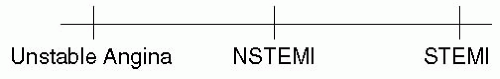
FIGURE 3-1 Spectrum of ACS.
Unstable Angina
Angina pectoris or ischemic symptoms with at least one of the following:
  • Occurring at rest or with minimal exertion and usually lasting >20 minutes (if not interrupted by nitroglycerin)
  • Severe pain of new onset (i.e., <1 month)
  • Occurring with a crescendo pattern (i.e., more severe, prolonged, or frequent than previously)
  • No evidence of myocardial infarction by biomarkers
Myocardial Infarction2
The typical rise and fall of troponin or creatinine kinase MB fraction (CK-MB) with at least one of the following:
  • Ischemic symptoms
  • Development of pathologic Q waves on the ECG
  • ST elevation, depression or evolutionary EKG changes consistent with ischemia
  • Post PCI (CK-MB 3× upper limit of normal)
Non-ST Segment Elevation MI (NSTEMI)
Criteria for MI in the absence of or with only transient ST elevation
ST Segment Elevation MI (STEMI)
Criteria for MI in the presence of persistent ST elevation indicative of myocardial injury
NQWMI/QWMI
The older literature makes reference to Q wave MI (QWMI) and Non-Q wave MI (NQWMI). The implication was that Q wave MI was associated with transmural infarction and Non-Q wave MI was limited to the subendocardium. Pathology studies have shown that Q waves are neither sensitive nor specific for transmural infarction. While there may be prognostic value in the identification of Q waves in patients with myocardial infarction, the modern terms STEMI and NSTEMI are preferred as they have more immediate implications with regard to acute management strategies (i.e., STEMI is triaged toward emergent reperfusion)
Causes of ST Elevation Other than AMI
Normal Variants
  • Normal male pattern: Often with 1-3 mm of concave sinus tachycardia (ST) elevation greatest lead V2
  • Early repolarization: Often with 1-3 mm of concave ST elevation, often with notching at the J point in V4
  • Variant ST elevation with T wave inversion: Seen often in young black men, convex ST elevation, T wave inversion, thought to represent persistent juvenile T wave inversion + early repol, may look like STEMI
Other Pathology
  • Left bundle branch block (LBBB): Typical pattern of LBBB present with anterior ST elevation
  • Pericarditis: Diffuse ST elevation and PR depression, PR elevation in AVR, the ratio of ST elevation: T wave height (> 0.25 measured using PR segment as the baseline in this case) in V6 is highly sensitive and specific for the diagnosis of pericarditis
  • Left ventricular hypertrophy with strain
  • Hyperkalemia: Downsloping ST elevation associated with wide QRS, peaked T waves
  • Pulmonary embolism: Sinus tachycardia, often anterior T wave inversions
  • Takotsubo cardiomyopathy (apical ballooning syndrome)
  • Brugada syndrome: Right bundle, downsloping ST elevation >2mm isolated to V1 and V2
DIFFERENTIAL DIAGNOSIS OF CHEST DISCOMFORT
TABLE 3-1 Immediately life-threatening causes (must consider these every time)

Disorder

History

Classic Findings

ACS

Substernal pressure with radiation to jaw, shoulder, arm, nausea, diaphoresis, often similar in character to previous angina. History of CAD, HTN, DM, hyperlipidemia, smoker, advanced age

Tachycardia, S4, new MR murmur or normal exam

Aortic dissection

Acute onset, tearing CP, radiation to back, may be associated with stroke symptoms. History of HTN, Marfan syndrome

Classic clinical ACS with normal ECG

May see ST elevation, classically inferior from RCA dissection

Widened mediastinum

Good positive predictors of the diagnosis: Pulse deficits and focal neuro findings with CP3

New AI murmur

Poor negative clinical predictors—if you think about it, exclude it

Pulmonary embolism

Acute onset, sharp, pleuritic CP, dyspnea, tachypnea. History/family history of DVT/PE/thrombophilia, malignancy, pelvic surgery, trauma, prolonged bed rest, travel, smoking, OCP

Always consider with hypoxia and a nl chest x-ray

ECG most commonly sinus tach, also anterior TWI, new RBBB, RAD

S1Q3T3 uncommon

Pneumothorax

Unilateral, sharp CP, pleuritic. History of trauma, asthma, COPD, mechanical ventilation, tall and skinny

Tracheal shift

Lucency of chest x-ray

Shock if under tension

TABLE 3-2 Urgent causes

Disorder

History

Classic Findings

Pericarditis

Sharp, localized, relief with sitting forward

Friction rub

Diffuse PR depression and/or ST elevation

PR elevation in AVR

ST elevation: T wave amplitude ratio in V6≥ 0.25 very high sensitivity/specificity for acute pericarditis (baseline is end of the PR segment)4

Pulmonary HTN

DOE, anginal-like pain (? RV ischemia), history-middle age women>men

Elevated JVP, loud P2, parasternal lift, S4

Stigmata of R sided HF

CXR with increased PA markings

See chapter on PH

Myocarditis

CP, fever, malaise, viral prodrome

Can elevate troponins and focal myocarditis can even cause WMAs

Esophageal rupture

Pleuritic CP, History of violent vomiting, esophagitis

Chest x-ray with mediastinal air

Hamman crunch on exam

Biliary tract disease

RUQ pain, postprandial, fever, history of gallstones, n/v

Murphy sign

Jaundice if common duct

Abnormal LFTs

Pancreatitis

Epigastric, radiates to back, alcoholic, History of pancreatitis or gallstones

LFTs in gallstone pancreatitis, Lipase

Pneumonia

Pleuritic, fever, cough, sputum production

Elevated WBC, fever

Rhonchi, rales

Chest x-ray with infiltrate

TABLE 3-3 Other causes

Disease

History

Classic findings

GERD

Long rest episodes with neg cardiac markers, burning, worse with recumbency, chocolate, caffeine, alleviated by antacids

Esophagitis on EGD

Promptly relieved with GI cocktail (be careful here)

PUD/Gastritis

Epigastric burning, worse after meals, caffeine, stress

Epigastric tenderness

Guaiac positive stools

Esophageal spasm

Substernal pain, aggravated with cold liquids

Often relieved with NTG (remember it’s smooth muscle too!)

Costochondritis or chest wall trauma

Localized pain, history of trauma or recent viral illness

Tender to palpation

Herpes Zoster

Unilateral local pain, immunosuppression, HIV

Pain often precedes rash

Make sure you look at the chest!

Anxiety

History of panic attacks, GAD, Depression, PTSD, recent traumatic event, on antidepressants/benzodiazepines

This has to be a diagnosis of exclusion

ACS, acute coronary syndrome; CAD, coronary artery disease; HTN, hypertension; DM, diabetes mellitus; MR, mitral regurgitation; CP, chest pain; RCA, right coronary artery; DVT, deep venous thrombosis; PE, pulmonary embolism; OCP, oral contraceptive pill; CXR, chest x-ray; TWI, t wave inversion; RBBB, right bundle branch block; RAD, right axis deviation; COPD, chronic obstructive pulmonary disease; AVR, aortic value replacement; JVP, jugular venous pressure; P2, pulmonic second sound; DOE, dyspnea on exertion; RV, right ventricle; PA, pulmonary artery; RUQ, right upper quadrant; LFT, liver function test; WBC, white blood count; GERD, gastroesophageal reflux disease; EGD, esophagogastroduodenoscopy; PUD, peptic ulcer disease; NTG, hitroglycerin; GAD, general anxiety disorder; PTSD, post-traumatic stress disorder.

Other Disorders Presenting with Chest Discomfort and ECG Changes (+/− Biomarker)
  • Takotsubo cardiomyopathy (apical ballooning syndrome)
  • Coronary vasospasm (Prinzmetal angina)
  • Coronary dissection
HISTORY
The history should be methodically taken and may be aided by remembering the “SOCRATES” mnemonic.
  • Severity: The 1-10 scale is useful, functional limitations should be sought
  • Onset: Be specific, can affect decisions about thrombolysis and PCI
  • Character: Pressure versus sharp, pleuritic, tearing (aortic dissection)
  • Radiation: Shoulder, jaw, arm, back
  • Associated symptoms: shortness of breath (SOB), nausea/vomiting (N/V), palpitations, syncope
  • Tempering factors: Rest, particular position (pericarditis—relief sitting forward)
  • Exacerbating factors: Exertion, emotional state versus particular position
  • Self assessment: Knowing the patient’s fears is important for many reasons
  • Also look for history of CAD, details of prior angiography, CABG or percutaneous coronary intervention (PCI), recent stress tests or echoes, other risk factors (DM, smoking, HTN, hyperlipidemia, cocaine use)
PHYSICAL EXAM
The physical exam in someone having an ACS may be entirely normal. Its purpose is to identify early complications of ACS as well as other possible etiologies of CP.
Look for:
  • Hypotension/hypertension
  • Arrhythmias
  • Tachypnea or hypoxia
  • Fever
  • Bilateral SBP difference >15 mm Hg (think of aortic dissection)
  • Asymmetric pulses and/or a focal neurologic exam (aortic dissection)
  • Signs of heart failure (elevated JVP, S3, crackles, hemoptysis)
  • New MR murmur (? papillary muscle ischemia)
  • A new S4 in a patient with active CP is highly suggestive of ischemia
  • Murmur of aortic stenosis, pulsus parvus et tardus
  • Abdominal tenderness (biliary tract/pancreatic disease)
  • Rectal exam—evaluate for melena or guaiac positive stool (? how aggressive to anticoagulate)
  • Rash on the chest (Zoster), make sure to examine the chest on hospital day 2, the pain often precedes the skin findings!
LABORATORY EXAMS AND IMAGING
Cardiac Biomarkers
  • Troponin I or T may be detectable as early as 2 hours after injury and CK-MB become detectable around 6 hours and are most sensitive at 10 hours
  • Myoglobin may rise somewhat sooner but is nonspecific
  • Must obtain serial tests q4-6 h for at least 10 hours after the start of CP to definitively r/o MI
  • False positive in heart failure, significant hypertension, PE
  • Look for typical rise and fall of enzymes in the context of CP and the ECG to make a diagnosis of NSTEMI
  • Heart failure can be responsible for persistent low-grade elevation of troponin thought to be from elevated LVEDP leading to subendocardial necrosis often with normal CKs
Additional Labs
  • WBC: Likely mildly elevated in AMI but may also establish infectious etiology for current state
  • Hemoglobin: Anemia contributes to patients presenting with shortness of breath or angina
  • Platelets: Severely depressed <50k levels may be a contraindication to PCI
  • Electrolytes: Monitor for potassium and calcium abnormalities which may alter the ECG, Evaluate for infectious etiologies, electrolyte abnormalities contributing to arrhythmias, biliary tract disease, or pancreatitis, if indicated
  • Creatinine: Usually elevated in patients with longstanding HTN and DM. Elevated creatinine is a relative contraindication to angiography because of the risk of renal failure secondary to contrast nephropathy
  • LFTs: Elevated in cases of hypotension due to shock or passive congestion due to elevated right-sided filling pressures. Also serum glutamic oxaloacetic transaminase/aspartate aminotransferase (SGOT/AST) can be elevated from an acute coronary syndrome as it is released from myocardial necrosis
  • Urine toxicology: Should be obtained for all young or at-risk patients with concerning chest pain (cocaine-induced MI can result from thrombosis in addition to vasospasm and therefore reperfusion may be indicated)
  • B-type natriuretic peptide (BNP): May be helpful in establishing the diagnosis of congestive heart failure (CHF) (see chapter on complications of AMI)
ECG
  • Should be obtained within 5 minutes of arrival within the emergency department
  • Make sure to label each ECG with the degree of pain
  • Comparison to an old ECG can be helpful
  • Repeat every 15 minutes ×2 if nondiagnostic, this has been shown to increase sensitivity
  • Dynamic T wave and ST changes should greatly increase your suspicion for ACS
  • Reoccurrence of CP should be re-evaluated with additional ECG recordings prior to giving NTG
  • Obtain right-sided leads in those with inferior distribution injury to evaluate for RV injury or infarct
  • Obtain posterior leads in patients with only anterior ST depression to r/o posterior wall STEMI
Chest X-ray
  • Assess for a widened mediastinum and always consider aortic dissection
  • Examine lung fields for evidence of a pneumonic process, pulmonary congestion, or pneumothorax
  • Assess ribs and soft tissues for evidence of trauma
  • Hypoxia/tachypnea with a clear CXR? Consider PE
Echo
  • Useful test for evaluating systolic function, regional WMAs, effusion, valvular lesions, and to assess for complications of MI
  • May be helpful in a high risk patient with ongoing CP and a nondiagnostic ECG or when the diagnosis of STEMI is in doubt
Helical CT of the Chest
  • Used to assess for proximal PE and aortic dissection and to assess for other noncardiac etiologies of CP such as pneumonia or pulmonary mass
  • Sensitivity and specificity are institutional and reader dependent
  • Consider MRI if patient has CRI or is high risk for contrast nephropathy
  • Different phase of contrast often needed for optimal evaluation of PE and aortic dissection
MANAGEMENT—NSTE ACS VERSUS STEMI
Differentiation of STEMI from non-ST-segment-elevation acute coronary syndrome (NSTE ACS) is critical to optimal management. These two entities have vastly different priorities as STEMI often involves a completely occluded artery and clinical data indicate that revascularization is both an emergency and that outcomes are tied to time to reperfusion. Management of NSTE ACS, both from a pathophysiologic and a clinical outcomes perspective focus on initial treatment with drugs that inhibit thrombosis and platelet aggregation usually in conjunction with a nonemergent early interventional approach and early adjunctive medical therapies. The major difference is the prioritization of treatments and the recognition of STEMI as a time-critical emergency.
TABLE 3-4 TIMI risk score (TRS) for UA/NSTEMI11

Score calculation (1 point each)

Score

D/MI/Revasc @ 14 days

Age ≥65

0-1

5%

≥3 risk factors for CAD

2

8%

Known CAD (>50% stenoses)

3

13%

ASA use in past 7 d

4

20%

≥2 episodes of angina in 24 h

5

26%

ST deviation ≥0.5 mm

6-7

41%

(+) cardiac enzymes

Add total points 0-7

INITIAL EVALUATION AND RISK STRATIFICATION
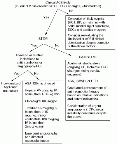
FIGURE 3-2 Approach to antithrombotic therapy in suspected ACS.
TABLE 3-5 UA/NSTEMI ACS risk stratification

High risk (≥1 of:)

Low risk (all of:)

History

Prolonged anginal CP or CP relieved only with maximal medical treatment

New CCS II-IV angina in the past 2 weeks but NO CP >20 min

ECG

ST ↓≥0.5 mm in two contiguous leads or

No ST ↓ or TWI

Deep TWIs >2mm excluding AVR and V1

Troponin

(+) Troponin

(−) Troponin

TIMI risk score

≥3

≤2

TABLE 3-6 Immediate NSTEMI ACS treatment strategies checklist

Low risk

High risk

ASA 162-325 mg5 (If allergy use Clopidogrel instead)

X

X

O2 (To keep SaO2>90%)

X

X

B-Blockers6

X

X

Lopressor 5 mg IVP q 5 min ×3 then 50 mg po q6h and titrate as tolerated. Harm likely in pts with significant CHF (see COMMITT CCS-2 below)

(Contraindicated HR <55, SBP <110mm Hg, PR >0.24 S or advanced heart block, mod-severe CHF, severe asthma, cocaine ACS. If actively wheezing, consider Diltiazem for significant tachycardia)

Nitroglycerin

X

X

SL, Paste, IV

(Contraindicated in severe AS, HOCM, RV infarct, and Viagra/Levitra/Cialis use)

Morphine

X

X

Prn pain, anxiety

K>4.0 meq/dL, Mg>2.0 meq/dL

X

X

Decreases risk of arrhythmias

DVT prophylaxis

X

X

(SQ heparin or LMWH prophylaxis for those not already on full anticoagulation)

Enoxaparin or UFH7

X

Enoxaparin 1 Mg/Kg SC q 12 hr, UFH may be favored if urgent cath

(If Cr > 2.5, CrCl < 30 ml/min, or weight > 150 kg use UFH, relative contraindication: significant anemia, low plts)

Clopidogrel8

X

600 mg po load then 75 mg daily

(Contraindicated with thrombocytopenia and caution with guaiac positive patients, caution if high likelihood of CABG, consult fellow/attending)

GPIIB/IIIA inhibitor9

X

(Start immediately for STEMI. In NSTEMI benefit probably only in troponin + patients, PCI, and greatest benefit in diabetics, consult fellow/attending for guidance)

High dose statin10

X

Lipitor 40-80 mg po daily or Simvastatin 40-80 mg po qhs (check baseline LFTs, lipid panel in am on all ACS patients)

ACE inhibitor before discharge for ↓ EF or HF

X

Captopril 6.25 mg po tid and titrate to goal 50 mg tid Ramipril 2.5 mg po and titrate to goal 5 mg po bid (Especially with anterior MI orEF, contraindicated if hypotensive, ARF orK+)

Early coronary angiography and reperfusion

X

(See chapter on angiography and PCI)

Adapted from the ACC/AHA 2002/2004 practice guidelines. (TIMI), thrombolysis in myocardial infarction

Only gold members can continue reading. Log In or Register to continue

Stay updated, free articles. Join our Telegram channel

Jul 16, 2016 | Posted by in CARDIOLOGY | Comments Off on Chest Pain and the Acute Coronary Syndromes

Full access? Get Clinical Tree

Get Clinical Tree app for offline access