CHEMICAL CONTROL OF BREATHING

9


CHEMICAL CONTROL OF BREATHING


Introduction


This chapter is only separated from the following one on neural control of breathing for your ease of understanding. All control of breathing is fundamentally neural. The sensory cells that detect changes in the external environment and the composition of the blood and cerebrospinal fluid, the central processors in the brain and the outputs that activate the muscles of breathing are all nerves.


A major difference between ‘neural control’, dealt with in the next chapter, and chemical control of breathing is the difference in timescale of their responses. Neural control responds in fractions of a second and changes the size and duration of individual breaths. Chemical control is normally much slower in its response, changing breathing minute by minute. In essence, chemical control determines minute ventilation, whereas neural control determines the most efficient pattern to achieve that ventilation with the minimum expenditure of work.


The ‘objective’ of respiration is homeostasis of arterial blood in terms of O2 and CO2 (which is closely related to arterial [H+]). This is achieved by matching ventilation to the metabolic activity of the body. This matching requires monitoring of the chemical composition of arterial blood, and the sensors which act as monitors are known as chemoreceptors.


Just as we have divided the subject of control of breathing into neural and chemical, so we can divide chemical control of breathing into sections in terms of the anatomical location of the sensors or, alternatively, what they are sensitive to. Those within the central nervous system are called central chemoreceptors and those outside peripheral chemoreceptors. Central chemoreceptors are most sensitive to excess CO2; peripheral chemoreceptors are most sensitive to lack of O2.


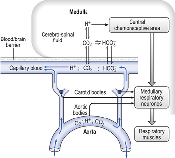
It is rare for excess CO2 or lack of O2 to occur alone: they usually occur together, and the whole chemoreceptor system is shown schematically in Figure 9.1.


Oxygen lack


The term for a lack of oxygen in any gas mixture or solution is hypoxia. Lack of O2 in arterial blood is termed hypoxaemia. Total absence of O2 is anoxia. It is very easy to change the amount of a gas in the arterial blood by utilizing the powerful gas-transporting properties of the lungs. Simply giving a subject a gas mixture to breathe will result in his or her arterial blood taking on the composition of that gas mixture within remarkably few breaths. The rate at which equilibrium is reached depends on the solubility of the gas in body fluids, and this has important consequences in anaesthesia. However, for the gases we are concerned with here equilibrium is approached within a few dozen breaths. The chemoreceptors that sense lack of arterial O2 are the carotid bodies and the aortic bodies. In humans it is the carotid bodies that are mainly responsible for the respiratory response. They are small (5.0 mm diameter) nodules of glomus tissue (Latin glomerus, a skein or ball of thread, i.e. a knot of capillaries) situated near the bifurcation of each common carotid artery. Unlike the carotid bodies, which mainly respond to Pao2, the aortic bodies are stimulated by reductions in arterial O2 content, e.g. carbon monoxide poisoning and anaemia affect them more. So it seems that the aortic bodies are sensitive to the total amount of O2 delivered to them, and the carotid bodies are sensitive to Pao2. The carotid bodies are situated close to the baroreceptor region of the carotid arteries, which help to regulate blood pressure, and are frequently confused with them. The carotid bodies are not baroreceptors.


Case 9.1   Chemical control of breathing: 1


Chronic obstructive pulmonary disease


Mrs Andrews is a 69-year-old lady who suffers from chronic obstructive pulmonary disease (COPD). This has been brought about by many years of heavy smoking – Mrs Andrews smokes 30 cigarettes per day and has done since she was a teenager. Mrs Andrews has a cough that is usually productive of white sputum. She often feels breathless and ‘wheezy’, and takes two bronchodilator drugs via an inhaler. She frequently suffers from chest infections that are usually treated with antibiotics by her doctor.


One winter, Mrs Andrews contracted a particularly severe chest infection. She had a cough productive of large volumes of green sputum and became very breathless indeed. Her own doctor decided to admit her to hospital for treatment.


In hospital, Mrs Andrews was found to be cyanosed and arterial blood gases indicated that she was hypoxic with a Pao2 of 6.2 kPa breathing. Her blood gases also indicated that her Paco2 was raised at 7.3 kPa. Initially, she was given oxygen to breathe. Although this resulted in the Pao2 increasing to 10.8 kPa, it also resulted in an increase in Paco2 to 8.4 kPa. At this stage, she was becoming very breathless and the effort of breathing was starting to exhaust her. The decision was taken to ventilate her lungs artificially while she received treatment for her infection and she was taken to the intensive care unit.


In this chapter we will consider:


Histology, embryology and anatomy of the carotid bodies


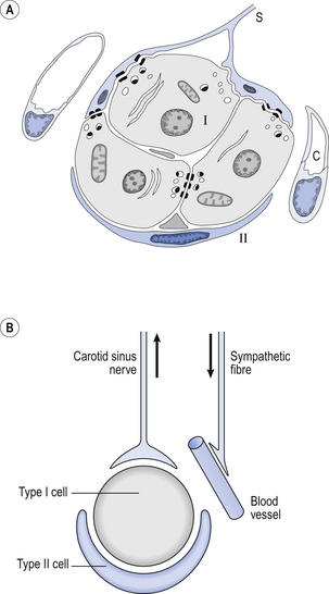
The function of the carotid bodies is related to their unusual structure. They have an extremely high metabolic rate (about three times that of the brain) but their rate of perfusion by blood from the carotid arteries is even higher: 10 times that which would be expected. This blood flows through capillaries (Fig. 9.2A and B) which surround the sensory elements (the glomus or type I cells) that monitor blood Po2. The type I cells seem to be supported by type II (sustentacular) cells, whose function is still not clear. The type I cells send their information to the brain via the carotid sinus nerve, a branch of the glossopharyngeal nerve (Fig. 9.2A and B), which also provides them with sympathetic and parasympathetic innervation. A separate supply of sympathetic fibres from the nearby superior cervical ganglion innervates the carotid bodies’ blood vessels.


The overall effect of this extensive sympathetic and parasympathetic supply to the carotid bodies is that their sensitivity can be altered by:


As far as these influences on neurotransmission from the chemoreceptor cells to the afferent sensory nerve endings is concerned it has become frustratingly obvious that, like many CNS synapses, there is a complex interplay of neuromodulators. However, the general consensus is that whereas sympathetic activity may mildly modulate carotid body function it does not have powerful effects on hypoxic ventilation. Dopamine, on the other hand, appears to be an important neuromodulator in the carotid body, inhibiting ventilatory responses to hypoxia.


The embryological origin of the carotid bodies provokes an interesting speculation. As a mammalian embryo develops its structure changes, resembling successive adult forms of more primitive species, starting with the most primitive and finally reaching mammalian form. This is the (now questionable) concept that ‘ontogony recapitulates phylogeny’. During our fish-like phase in the womb those structures that are going to become our O2-sensitive carotid bodies are represented by the gill arches of the fish-like embryo. It is the gills of fish that are their sensors of O2 lack. It is therefore postulated that our carotid bodies are a residue of the mechanism by which our fishy ancestors detected O2 lack in their watery environment.


Hypoxic stimulation


Activity in the carotid bodies is measured experimentally as the frequency of discharge of action potentials in the carotid sinus nerve. Increased activity expresses itself in the whole animal as an increase in ventilation. Hypoxia stimulates peripheral chemoreceptors, which is unusual, as the activity of almost all other organs is depressed by it.


During eupnea under normoxic conditions most of the drive to breathe comes from central chemoreceptors, and also neural mechanisms associated with wakefulness. Evidence that the peripheral chemoreceptors provide some drive to breathe comes from the observation that in patients who have been subjected to carotid body denervation arterial Pco2 is elevated by up to 0.8 kPa.


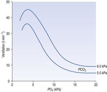
The effect of decreasing a subject’s arterial Po2 by giving them increasingly hypoxic gas to breathe is shown in Figure 9.3.


It can be seen that Pao2 must be reduced considerably (to about half normal) before breathing is stimulated, and that very low partial pressures of O2 depress breathing.


Hypercapnic stimulation


Increased levels of arterial CO2 (hypercapnia) also stimulate peripheral chemoreceptor activity, probably by increasing [H+] within the glomus cells, in the same way as increased extracellular acidity increases chemoreceptor activity and breathing.


What is the actual physiological stimulus to the carotid bodies? It is difficult to see how the absence of something, in this case O2, can be a stimulus. A number of different observations combine to give us a clue:


1. Chemoreceptors have a very high metabolic rate and so rapidly use up O2 supplied to them.


2. They have a very high blood flow, gram for gram 40 times that of the brain.


3. Pao2 must be reduced considerably before there is stimulation of breathing, but then the increase is large.


4. Increasing Pao2 above normal (13 kPa) by inhaling O2-rich mixtures only produces a small reduction in breathing by depressing chemoreceptor activity.


5. Increasing arterial [H+] does not have a great effect on central chemoreceptors but stimulates peripheral chemoreceptors.


6. Peripheral chemoreceptors are much less sensitive to increases in Paco2

Stay updated, free articles. Join our Telegram channel

Jun 18, 2016 | Posted by in RESPIRATORY | Comments Off on CHEMICAL CONTROL OF BREATHING

Full access? Get Clinical Tree

Get Clinical Tree app for offline access