Changes in the Pharmacokinetics and Pharmacodynamics of Drugs Administered during Cardiopulmonary Bypass


Changes in the Pharmacokinetics and Pharmacodynamics of Drugs Administered during Cardiopulmonary Bypass


Richard I. Hall

Derek J. Roberts


Cardiac surgery may be performed with or without cardiopulmonary bypass (CPB) (1). When utilized, practitioners should be aware that CPB profoundly affects the way drugs are distributed and cleared by the body (i.e., drug pharmacokinetics [PK]) and how they interact with the body to produce their effects (i.e., pharmacodynamics [PD]) (2). This chapter reviews some basic pharmacokinetic and pharmacodynamic concepts and then describes the role that CPB (and the systemic inflammatory response that it generates) may play in altering the pharmacokinetics and pharmacodynamics of drugs administered during cardiac surgery. An understanding of the above may allow for practitioners to explain apparent anomalies in drug action. This may include enhanced intravenous anesthetic effect in the presence of hemodilution during CPB (3,4,5,6,7,8,9,10,11) or the potential for development of awareness during CPB under anesthesia due to insufficient volatile anesthetic drug administration (12,13,14).


BASIC PRINCIPLES AND DEFINITION OF TERMS

To understand how CPB may alter the effect of drugs, one must first understand some basic pharmacokinetic and pharmacodynamic principles.


Pharmacokinetics

Pharmacokinetics is defined as the mathematical description of the processes through which a drug is handled once introduced into the body, that is, what the body does to the drug. Because the vast majority of drugs given during cardiac surgery are administered intravenously, this discussion will be primarily limited to a description of the pharmacokinetic principles involved in describing the fate of a drug administered during intravenous drug administration.

Following injection of a single intravenous dose of a drug (e.g., induction of anesthesia), a number of processes are initiated that serve to reduce drug concentrations. The drug is delivered to and taken up by tissues within the body—a process known as distribution. Distribution to highly perfused tissues such as the brain, heart, lungs, liver, and kidneys occurs first. Tissue uptake at this stage is variable, depending on factors such as protein binding (typically decreased uptake with increased plasma protein binding) and the lipid solubility of the drug (typically increased uptake with increased lipid solubility). Thereafter, distribution occurs into less well perfused tissues such as muscle and fat. As the drug is delivered to organs such as the liver, kidneys, and lungs, elimination by biotransformation and excretion occurs. Elimination may be influenced by age (15), gender (16), disease (17), and CPB (18,19). For most drugs employed during cardiac surgery, elimination occurs as a constant fraction of drug remaining in the body per unit time. This is known as first-order kinetics.

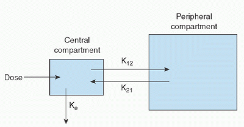
Various mathematical models have been developed to quantify what happens to a drug once it is introduced into the body. For the high-potency opioids, during cardiac surgery, a simple two- (20) or three (21)-compartmental analysis has been shown to adequately serve for clinical purposes. Figure 10.1 depicts a two-compartment model. Following drug injection, distribution occurs within a central compartment (blood) and to the peripheral compartments (tissues). Transfer of the drug between the central and peripheral compartments can be described by appropriate rate constants
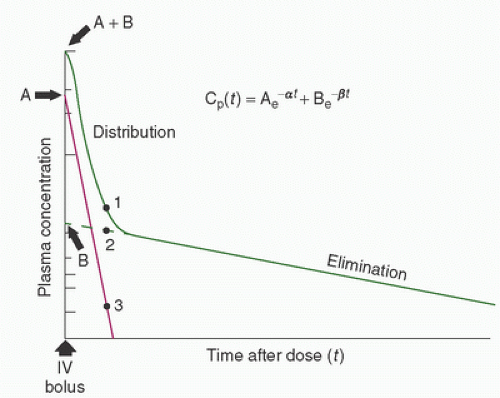
(Fig. 10.1). Elimination occurs from the central compartment and can be described by the elimination rate constant. By measuring plasma concentrations over a time period from the injection of the drug, it is possible to describe the concentration-versus-time profile for the drug (Fig. 10.2) (22). Distribution and elimination phases can be determined and a mathematical description (model) of the change in drug concentration versus time can be developed.


FIGURE 10.1. A two-compartment pharmacokinetic model illustrating the distribution of a drug within a central compartment (blood) and peripheral compartment (tissues), and its ultimate biotransformation and elimination from the body. K12 and K21 are first-order rate constants for transfer of drug between the peripheral and central compartments, whereas Ke is the elimination rate constant. (From Hall RI, Thomas BL, Hug CC Jr. Pharmacokinetics and pharmacodynamics during cardiac surgery and cardiopulmonary bypass. In: Mora CT, ed. Cardiopulmonary bypass: Principles and techniques of extracorporeal circulation. New York, NY: Springer-Verlag, 1995:56.)

More sophisticated compartmental models can also be developed. For example, a three-compartment model that characterizes distribution to both highly perfused tissues and less highly perfused tissues and also describes the elimination phase can be developed. Other models account for additional pharmacokinetic details, including the very early distribution phase (23), tissue distribution (24), gender, weight, hemodilution (25), and institution of CPB (26). Strategies exist to determine which mathematical model best describes the observed concentration-versus-time profile for any drug administered (27). Derivation of the rate constants then allows for the development of computer programs designed to produce continuous infusions of drugs (e.g., computer-assisted continuous infusion [CACI]) at rates that maintain a stable targeted plasma concentration (21,25,28,29,30,31,32,33). Such information can be used to investigate concentration-versus-effect relations (10,34,35,36,37,38) and the influence of drug interactions (32,39,40,41,42). Characterization of concentration-versus-time profiles for intravenously administered drugs also allows derivation of other pharmacokinetic parameters such as the volume of distribution (Vd), clearance (Cl), and elimination half-time (t1/2β).


FIGURE 10.2. Plasma [log] concentration-versus-time curve for a hypothetical drug after a single intravenous dose. The curve (A + B) is the sum of the contributions from the rapid distribution (A) phase and the slow elimination (B) phase to the logarithmic decline in concentration after a bolus dose. The concentration at any time is given by the equation Cp(t) = Ae-at + Bet, where Cp(t) is the drug concentration in plasma at time t; A, constant determined from the Y-axis intercept (time = 0) of the distribution portion of the log concentration-versus-time curve, derived by subtracting the contribution of the (constant, first order) elimination phase of the curve; α, slope of the log concentration-versus-time curve of the distribution phase, derived by subtracting the contribution due to elimination; B, constant determined from the Y-axis intercept (time = 0) of the elimination phase of the log concentration-versus-time curve; β, slope of the log concentration-versus-time curve of the elimination phase. (From Hall RI, Thomas BL, Hug CC Jr. Pharmacokinetics and pharmacodynamics during cardiac surgery and cardiopulmonary bypass. In: Mora CT, ed. Cardiopulmonary bypass. Principles and techniques of extracorporeal circulation. New York: Springer-Verlag, 1995:56.)

Table 10.1 provides a list of terms commonly employed in the description of a drug’s pharmacokinetic properties (43).

Volume of distribution (Vd) is defined as that volume of fluid into which a drug would be administered in order to produce the observed concentration of drug in plasma. It does not correspond directly to any particular tissue compartment but is rather useful in predicting drug concentrations based on pharmacokinetic parameters. Vd is used to characterize the total volume of distribution, whereas Vdc describes the volume of the central compartment, or the initial volume of distribution (also termed Vi). Vdss describes the volume of distribution when steady-state plasma concentrations of a drug are achieved. Clearance (Cl) refers to the removal of a drug from the body, usually by way of the central compartment, and is expressed as the volume of blood completely cleared of the drug per unit of time. Elimination half-time (t1/2β) is the time required for the concentration of a drug in plasma to decrease by half. It can be determined by examining the elimination portion of the concentration-versus-time curve, or by substituting the relevant parameters in the following equation:


Similarly, distribution half-times (t1/2ρ, t1/2α) can be determined by examining the distribution phase(s) of the concentration-versus-time curve. Drugs with short elimination half-times are characterized by small volumes of distribution and/or rapid clearance. Drugs that have a long elimination half-time tend to be highly lipid soluble (most anesthetic agents) with a large volume of distribution and/or slow rate of elimination.

The degree to which drug effect terminates depends on the rapidity of drug redistribution to the central compartment once the injection stops, and the capacity of the elimination processes to clear the drug. Although this concept is important following the injection of a single dose, it is also important when drugs are given by continuous infusion or in repeated doses. If drug administration exceeds the body’s ability to clear it, drug accumulation will occur, leading to prolonged drug effect. At times, drug administration may completely saturate clearance mechanisms (e.g., excess alcohol ingestion), leading to a situation where clearance is no longer a function of drug level in the plasma and instead occurs at a relatively constant rate (so-called zero-order kinetics). More commonly,
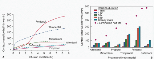
as drug administration continues, there is accumulation of drug in tissues over time, which increases with the duration of drug infusion. On termination of the infusion, offset of drug effect then depends on redistribution of the drug out of tissues (greater with longer infusions) back into the central compartment as well as the rate of elimination. Thus, when a drug has accumulated in peripheral tissues, reliance on the elimination half-time will not adequately predict termination of drug effect because it does not take into account the role of redistribution. This has led to the introduction of the term context-sensitive half-time as a better description of the phenomenon of increased duration of drug effect with increased drug infusion time (Fig. 10.3) (44).


TABLE 10.1. Definition of basic pharmacokinetic parameters


Parameter


Abbreviation


Definition


Area under the curve


AUC


Area under the concentration vs. time curve, which is commonly bounded by a time period, e.g., 0-4 h.


Bioavailability


F


The fraction of an administered dose which reaches the systemic circulation unchanged. Typically applied in reference to oral drug administration as the F of an intravenous drug is, by definition, equal to 1.


Clearance


Cl


The volume of blood cleared completely of drug per unit of time.


Elimination rate constant


Ke


The rate at which a drug is eliminated from the body per unit of time. Ke is inversely proportional to the elimination half-life of the drug.


Extraction ratio


ER


The percentage of medication removed from the blood as it passes through the eliminating organ. The extraction ratio depends not only on the blood flow rate but also on the free fraction of drug and the intrinsic ability of the organ to eliminate the drug. Typically used to describe how the liver handles a given drug.


Half-life


t1/2


The amount of time required for the drug concentration to decrease by 50%. The half-life may be determined for both the distribution (e.g., t1/2α) and elimination (e.g., t1/2β) phases of drug handling.


Plasma protein binding


Fu


The process by which a drug binds to proteins in the plasma until an equilibrium is established between the fraction bound (Fb) and the fraction unbound (Fu). Only the Fu is available to distribute, exert its pharmacologic effect, and be metabolized and eliminated.


Steady state


A condition where the rate of drug administration is equal to the rate of elimination. Steady state is generally reached after four or five half-lives of drug administration. Once achieved, drug elimination is generally considered to be complete after four or five half-lives once drug administration has been terminated.


Time to maximum concentration


TMAX


The time required to reach maximal blood concentration after drug administration.


Volume of distribution


Vd


The apparent or theoretical volume into which the drug distributes that relates the plasma concentration to the administered dose.


Equilibration rate constant


Ke0


Rate constant for equilibration at the site of drug effect.


Modified from Smith BS, Yogaratnam D, Levasseur-Franklin KE, et al. Introduction to pharmacokinetics in the critically ill. Chest 2012;141(5):1327-1336.


Computer-driven infusions of drugs use the pharmacokinetic parameters previously derived from concentration-versus-time profiles to set their infusion rates. The accuracy of these infusions in achieving the desired plasma concentrations therefore depends on the accuracy of the initial parameter estimates. Errors in these parameters can occur due to a variety of reasons, including number of drug measurement points (too few), duration of drug measurements (too short), sensitivity of the drug assay employed (too low), and nature
of the population studied (e.g., young, healthy men versus elderly women with congestive heart failure [CHF]). Drug in the blood exists in several forms, including that which is free (active), bound to plasma proteins (e.g., albumin, and therefore subject to changes in plasma protein concentrations), or sequestered in red blood cells. CPB has the potential to alter all of these factors, which makes the description of pharmacokinetic parameters during CPB problematic.


FIGURE 10.3. A: Context-sensitive half-times as a function of infusion duration for each pharmacokinetic model simulated. Solid and dashed lines are used to permit overlapping lines to be distinguished. B: Context-sensitive half-times (bars) redrawn from (A) for each pharmacokinetic model after terminating a 1-minute, 1-hour, 3-hour, 8-hour, or infinitely long (i.e., to steady state) computer-designed infusion designed to instantaneously achieve and maintain a target concentration shown relative to the elimination half-life (dots) computed for each model. (From Hughes MA, Glass PSA, Jacobs Jr. Context-sensitive half-time in multicompartment pharmacokinetic models for intravenous anesthetic drugs. Anesthesiology 1992;76:336.)

Where infusions are administered at constant rates (so-called zero-order infusions), drug accumulation over time is likely. To prevent drug accumulation, adjustment of infusion rates according to patient response is therefore strongly suggested (20). This should maintain plasma drug concentration in the lower therapeutic range and permit optimal reduction in concentration (and termination of drug effect) once the infusion is terminated.

Termination of drug effect depends highly on clearance mechanisms. For most drugs, this involves some degree of liver metabolism and/or renal excretion. Lipophilic drugs are metabolized in the liver in either one or two phases, which need not occur sequentially. Phase I reactions convert lipophilic drugs to more water-soluble compounds through oxidation, reduction, or hydrolytic reactions. Oxidation-reduction reactions occur in the endoplasmic reticulum and are frequently mediated by the cytochrome P-450 superfamily of mixed-function oxidases. These enzymes exist in a number of isoforms, each of which has a separate, but somewhat overlapping, list of particular drug and xenobiotic substrates (45). The cytochrome P-450 enzymes are regulated by gene transcription, and their activities can be modified by drugs and disease processes (46,47,48) as a result of enzyme induction (e.g., rifampin) (48) or inhibition (e.g., erythromycin (49), propofol (47), and fluconazole (50)). Their activities are also affected by genetic predisposition (e.g., heterogeneity in the ability to metabolize certain drugs (51)). Phase II differs from phase I reactions as they couple the drug (or its metabolites) to an endogenous substrate such as sulfate, acetate, or glucuronide to form a highly polar, water-soluble compound that is more easily excreted (52).

The ability of the liver to metabolize a drug in the absence of limitations imposed by hepatic blood flow or drug-protein binding is termed intrinsic hepatic clearance (53). The hepatic extraction ratio is the fraction of a drug contained in hepatic arterial blood that is removed as it passes through the liver. These two concepts are related by the following equation:


where Clhepatic = hepatic clearance rate of a drug, Q = liver blood flow, Cli = intrinsic hepatic clearance, Ca = arterial drug concentration, Cv = venous drug concentration, and E = hepatic extraction ratio.

Drugs with a low extraction ratio (e.g., diazepam (54)) depend on hepatic metabolism for their elimination and are much more affected by changes in protein binding and the liver’s ability to metabolize drugs (e.g., through induction or inhibition of cytochrome P-450 enzymes) than by changes in liver blood flow. In contrast, the metabolism of drugs with a moderate (e.g., alfentanil (55)) or a high extraction ratio (e.g., sufentanil (56) and propofol (8)) may be critically affected by changes in blood flow in the liver.

For drugs cleared by the kidneys, excretion depends on renal blood flow, glomerular filtration rate, tubular secretion, and reabsorption (57). When excreted by filtration (e.g., mannitol (58)), the rate will depend on the plasma concentration and renal blood flow. Drugs excreted by tubular processes (tubular secretion and reabsorption, e.g., cefazolin (59)) may be subject to saturation of active transport processes.


Pharmacodynamics

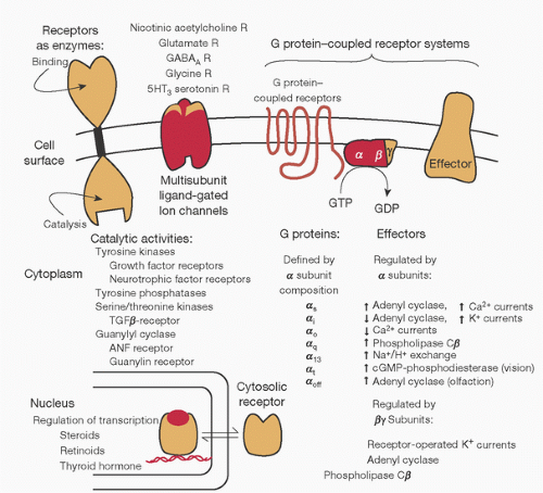
Pharmacodynamics describes how a drug interacts with the body to produce changes in patient physiology. Most drugs produce these effects by interaction with a specific receptor, that is, the macromolecular component of the organism with which the drug interacts through a lock-and-key or other type of mechanism (Fig. 10.4) (60). Activation of the receptor leads to changes intracellularly, often through secondary messengers, which in turn leads to changes in cell function (e.g., muscle contraction). Although other types exist (e.g., nucleic acids), receptors are most often proteins. Proteins that serve as receptors for endogenous ligands are particularly important because drugs that interact with these receptors produce physiologic effects mimicking those in nature (61,62,63,64). Drugs with this mechanism of action are referred to as agonists. In contrast, drugs that possess no intrinsic pharmacologic activity, but bind to receptors and interfere with binding of endogenous ligands, are termed antagonists.

Whether a drug acts as an agonist or antagonist depends on its structure, and this aspect is exploited in drug design. The degree to which a compound can mimic the effect of the endogenous ligand is also a function of receptor number, receptor affinity for the drug, and the drug concentration to which the receptor is exposed.


FIGURE 10.4. Structural motifs of physiologic receptors and their relation to signaling pathways. Schematic diagram of the diversity of mechanisms for control of cell function by receptors for endogenous agents acting through the cell surface or in the nucleus. (From Ross EM. Pharmacodynamics. Mechanisms of drug action and the relationship between drug concentration and effect. In: Hardman JG, Limbird LE, eds. Goodman and Gilman’s the pharmacological basis of therapeutics. 9th ed. Montreal, QC: McGraw-Hill, 1996:32.)


Receptor Types and Functions

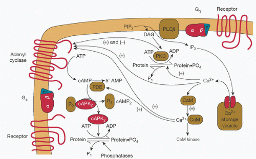
Receptors serve two functions: to bind the appropriate ligand and, following that, to propagate the regulatory signal into the target cell. This has led to the functional localization of two regions within the receptor—a ligand-binding domain and an effector domain. Receptor effects may be produced by action directly on its cellular target(s), effector proteins, or may be conveyed to other cellular targets by intermediary cellular molecules termed transducers (Fig. 10.4) (60). The combination of the receptor, its target proteins, and transducers constitutes the signal transduction pathway. In some cases, the effector protein may cause activation of another signaling pathway through secondary messengers (Fig. 10.5) (60).


FIGURE 10.5. Interactions between the second messengers cyclic adenosine monophosphate (cAMP) and Ca2+. Generation of second messengers cAMP and Ca2+ permits distribution of cell-surface regulatory input into the cell interior, amplification of the initial signal, and opportunities for synergistic or antagonistic regulation of other signaling pathways. PIP2, phosphatidylinositol 4,5-biophosphate; DAG, diacylglycerol; IP3, 1,4,5-inositol triphosphate; CaM, calmodulin; R2, regulatory subunits of cyclic AMP-dependent protein kinase, which bind cyclic AMP; cAPK2, catalytic subunits of cyclic AMP-dependent protein kinase; PKC, protein kinase C, activated by DAG and Ca2+. (From Ross EM. Pharmacodynamics. Mechanisms of drug action and the relation between drug concentration and effect. In: Hardman JG, Limbird LE, eds. Goodman and Gilman’s the pharmacological basis of therapeutics. 9th ed. Montreal, QC: McGraw-Hill, 1996:34.)

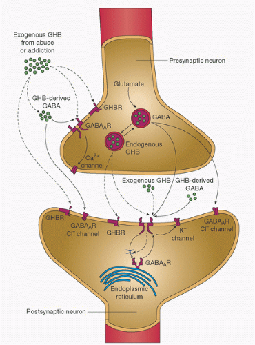
Families of receptors mediating a variety of functions have been characterized. Receptors for neurotransmitters (and exogenously administered drugs) are frequently in the form of an agonist-regulated, ion-selective channel in the plasma membrane, termed a ligand-gated ion channel (Fig. 10.6) (65). Stimulation of the receptor leads to changes in ion flux across the cell membrane, with subsequent alteration in the cell membrane potential or the cell’s ionic composition. Receptors in this group include nicotinic cholinergic receptors (site of skeletal muscle relaxant activity) (66) and the γ-aminobutyric acid type A (GABAA) receptor (which also serves as the receptor for benzodiazepines, propofol, and barbiturates) (67).

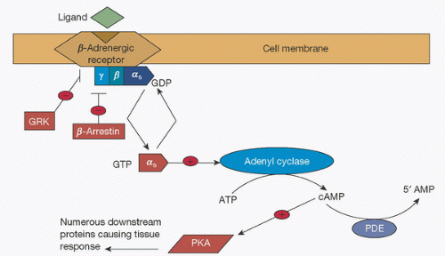
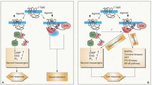
Many receptors (including opioid receptors (68)) are G-protein-coupled receptors (69). Occupation of the receptor by its ligand initiates binding of guanosine triphosphate (GTP) to specific G-proteins on the inner membrane surface, causing changes in the conformation of the G-protein complex and consequent signal transduction to specific effectors often mediated by second messenger enzymes such as adenyl cyclase and phospholipases A2, C1, and D. Effectors include channels specific for Na+, K+, and Ca2+ conductance and certain transport and regulatory proteins. The G-protein receptor has been well characterized and consists of seven α-helical segments spanning the cell plasma membrane (70). Ligand binding changes the conformation of α, β, and γ heterotrimetic G-protein-coupled receptor polypeptides on the inner surface of the plasma membrane (71). When receptor activation occurs, GTP binds to these subunits, resulting in disassociation of the a subunit from the βγ subunit such that interaction with the effector occurs (Fig. 10.7) (72). The βγ subunit may also interact with and influence effector activity. Termination of signal transmission occurs when G-protein-coupled receptor kinases (GRKs) phosphorylate the activated receptor, which leads to recruitment of β-arrestins and subsequent receptor desensitization. β-Arrestins may also serve as signal transducers (Fig. 10.8) (73). After CPB, GRK activity is reduced (74). Examples of G-protein-coupled receptors and their second messenger systems are given in Table 10.2 (69).

G-protein receptors are subject to regulatory and homeostatic controls. As an example, continuous stimulation of a receptor by an agonist (ligand) may result in a reduced effect as a result of processes known as desensitization, endocytosis, or downregulation. Desensitization is defined as any process that alters the functional coupling of a receptor to its G-protein/second messenger signaling pathway. Endocytosis is defined as the translocation of receptors from the cell surface to an intracellular compartment. Lastly, downregulation is defined as any process that decreases the number of ligand-binding sites (64,70).


FIGURE 10.6. Synthesis and release of γ-hydroxybutyric acid (GHB) and γ-aminobutyric acid (GABA) at synapses—an example of a ligand-gated ion channel. The diagram shows the presynaptic and postsynaptic effects of endogenously released GHB (as indicated by dashed arrows) and (GABA) (as indicated by solid arrows) and the effects of exogenously administered GHB, as in abuse and addiction. GABA is synthesized from glutamate in inhibitory neurons and in turn gives rise to GHB. Both GHB and GABA are released upon depolarization of the GABA-releasing (GABAergic) presynaptic neuron. GABA, in forms that are either endogenous or derived from exogenously administered GHB, acts on GABAA and GABAB receptors (GABAAR and GABABR, respectively). GABAA receptors are ionotropic and, when activated by GABA, cause fast postsynaptic inhibition by the efflux of chloride ions (Cl-). GABAB receptors are metabotropic and, when activated by either GABA or high concentrations of GHB, induce slow postsynaptic inhibition by activating outward potassium (K+) currents. Presynaptic GABAB autoreceptors—when activated by GHB, GABA, or both—reduce the release of GABA by suppressing the influx of calcium (Ca2+). Both endogenous and exogenous forms of GHB have a dual action on the GHB receptor (GHBR) and the GABAB receptor. GHB that binds with high affinity to the presynaptic GHB receptor decreases the release of GABA; GHB that binds to a low-affinity site on the GABAB receptor increases activation of cell-surface receptors by inhibiting constitutive and agonist-induced endocytosis. The result is enhancement of GHB function mediated by GABAB receptors, with a greater effect on presynaptic inhibition than on postsynaptic inhibition. (From Snead OC III, Gibson KM. γ-Hydroxybutyric acid. N Engl J Med 2005;352:26;2724.)


FIGURE 10.7. G-protein-coupled receptors. Schematic of signal transduction cascade for receptors coupling to G-protein [α]s subunit. ATP, adenosine triphosphate; cAMP, cyclic adenosine monophosphate; PDE, phospho-diesterase; PKA, protein kinase A; GRK, G-protein receptor kinase; GDP, guanosine diphosphate; GTP, guanosine triphosphate. (From Johnson JA, Lima JJ. Drug receptor polymorphisms and pharmacogenetics: current status and challenges. Pharmacogenetics 2003;13(9):531.)


FIGURE 10.8. Signal transduction by seven transmembrane receptors. A: Classical paradigm. The active form of the receptor (R*) stimulates heterotrimeric G-proteins and is rapidly phosphorylated by GRKs, which leads to β-arrestin recruitment. The receptor is thereby desensitized, and the signaling is stalled. B: New paradigm. β-Arrestins not only mediate desensitization of G-protein-signaling but also act as signal transducers themselves. TMR, transmembrane receptor; cAMP, cyclic adenosine monophosphate; DAG, diacylglycerol; IP3, 1,4,5-inositol triphosphate; Gα,γ,β, trimeric G-protein subunits; GRK, G-protein-coupled receptor kinases; MAPKs, mitogen-activated protein kinases; AKT, protein kinase B pathway; PI3, phosphatidylinositol-3-kinase pathway. (From Lefkowitz RJ, Shenoy SK. Transduction of receptor signals by β-arrestins. Science 2005;308:513.)


TABLE 10.2. Examples of G-protein-coupled receptors and the intracellular second messengers they generate


Receptor


Second messenger


Adrenoceptors


α1A/1B/1C


IP3+


α2A/2B/2C


cAMP-


β1/2/3


cAMP+


Bradykinin (B1-3)


IP3+


Calcitonin gene-related peptide


cAMP+


Dopamine (D1-5)


IP3+/cAMP-


Glutamate (metabotropic)


IP3+


Histamine


cAMP+/IP3+


5-HT (1A-1D/2/3/4)


cAMP-/cAMP+/IP3+


Muscarinic (M1-5)


IP3+/cAMP-


Opioid (µ/δ/κ)


cAMP-


Vasopressin (V1A,1B/2)


IP3+/cAMP+


cAMP+, receptor stimulates generation of cAMP; cAMP-, receptor inhibits generation of cAMP; IP3+, receptor stimulates generation of IP3; cAMP, cyclic adenosine monophosphate; IP3, inositol(1,4,5) triphosphate; HT, hydroxytryptamine. Reprinted from Lambert DG. Signal transduction: G proteins and second messengers. Br J Anaesth 1993:71;86-95.


Tolerance is a phenomenon whereby an increased amount of drug is required to produce the same level of pharmacologic effect after repeated use of the drug. The mechanism by which tolerance occurs is gradually being elucidated but is thought to represent a complex multifaceted process involving multiple regulatory processes at the cellular and neural circuit levels (75). High-potency opioids (e.g., fentanyl, sufentanil) with substantial intrinsic receptor affinity appear to produce tolerance faster than lower-potency opioids such as buprenorphine.

While the term tolerance is usually used to describe a loss of drug efficacy over hours to days, desensitization involves a more rapid loss of receptor activity. Desensitization can occur within a short time frame and lasts a short period of time (˜1 hour) in the absence of continued receptor stimulation. The mechanism appears to be multifactorial and includes phosphorylation of activated receptors, which results in their binding with a group of intracytoplasmic proteins termed arrestins (75,76). This binding causes an uncoupling of receptors from G-proteins and receptor desensitization. Once bound, the receptors may be dephosphorylated and returned to the cell surface, or be degraded by lysosomes (77). In the presence of an antagonist, the number of cell-surface receptors may also increase (78).

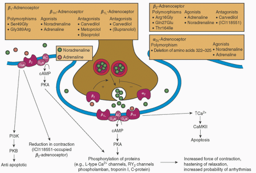
Traditionally, receptors have been classified by their physiologic effects and relative potencies. Examples include muscarinic versus nicotinic cholinergic receptors (79), α and βadrenergic receptors (80), and µ (mu), κ (kappa), and δ (delta) opioid receptors (81). Subtypes of receptors exist, for example, β1 and β2, and are targets for drug-selective effects (Fig. 10.9) (80). Molecular cloning techniques have allowed tissue-specific receptor subtypes to be identified and localized (61,64).

Soluble DNA-binding proteins that regulate transcription of specific genes serve as the receptor for steroid hormones, thyroid hormone, vitamin D, and the retinoids. They are part of a larger family of transcription factors that are regulated by phosphorylation, association with other protein factors, and/or by binding to metabolites or cellular regulatory ligands (82). Glucocorticoid receptors bind circulating adrenal steroids and are translocated into the cell nucleus, where they bind to glucose response elements to activate genes that encode anti-inflammatory proteins (83). The receptor is composed of three domains: a hormone binding region near the carboxyl terminus; a central region that interacts with nuclear DNA to activate or inhibit gene transcription (which, for glucocorticoids, is termed the “glucocorticoid-responsive element”); and an amino-terminal region whose function is not well defined (84).


Second Messenger Systems

Transduction of the signal from the receptor to the intracellular effector is often mediated by second messenger systems. These systems are relatively few in number. However, they affect the activity of many pathways, and receptor binding of a ligand may influence second messenger systems by altering the messenger’s function through activation (Gs) or inhibition (Gi) of G-proteins (Fig. 10.10) (85).

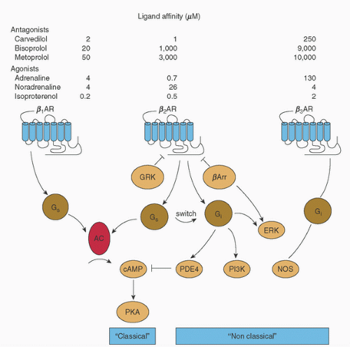
Cyclic adenosine monophosphate (cAMP) exemplifies the second messenger function. cAMP is synthesized by adenyl cyclase in response to receptor activation. Stimulation of adenyl cyclase activity is mediated by Gs and inhibited by Gi proteins (Fig. 10.11) (86). Its activation results in phosphorylation of phosphorylase kinase and downstream cellular responses, which may include glycogenolysis (87) or muscle contraction (86,88). Termination of cAMP activity is by targeted hydrolysis—a process catalyzed by several phosphodiesterases.

Intracellular Ca2+ serves as another second messenger (89). Intracellular calcium concentrations are controlled by
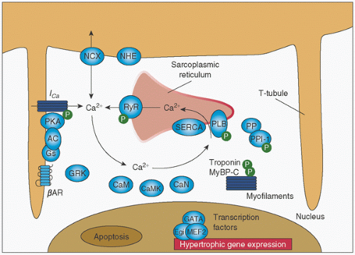
regulation of several different Ca2+-specific channels in the plasma membrane and by its release from intracellular storage sites (Fig. 10.12) (69,86,89). Ca2+-dependent ion channels are opened by electrical depolarization, phosphorylation by a cAMP-dependent protein kinase, and by Gs, K+, and Ca2+ itself (89). Opening of the channel may be inhibited by other proteins (e.g., Gi).


FIGURE 10.9. Features of the cardiac sympathetic nervous system. (—)-Noradrenaline released from sympathetic nerve terminals is complemented by circulating (—)-adrenaline. The release of (—)-noradrenaline is modulated by facilitatory, prejunctional β2-adrenoceptors and autoinhibitory, prejunctional α2-adrenoceptors. A deletion polymorphism of the autoinhibitory α2c-adrenoceptor reduces its function and leads to heightened noradrenaline release from prejunctional nerve terminals. In heart failure, the activity of the sympathetic nervous system increases, with a consequent increase in the plasma concentration of (—)-noradrenaline. (—)-Noradrenaline activates postjunctional β1-adrenoceptors that couple to the Gsα-cAMP pathway. Gsα activates adenyl cyclase (AC), which catalyzes the formation of cAMP. cAMP, in turn, activates cAMP-dependent protein kinase (PKA). PKA phosphorylates several proteins that contribute to increased force of contraction and hastening of relaxation. There are two forms of β1-adrenoceptors, β1H and β1L. In the human heart, β2-adrenoceptors also couple to the Gsα-cAMP pathway. Recently, it has been demonstrated that β1- and β2-adrenoceptors couple to additional signaling pathways that are of interest in heart disease. These include β2-adrenoceptor stimulation of Giα signaling pathways. Agonist-activated β2-adrenoceptor-Giα signaling has putative antiapoptotic effects through phosphatidylinositol 3-kinase (PI3K)-protein kinase B (PKB) signaling. The β2-adrenoceptor antagonist ICI118551 reduces human ventricular myocyte maximal shortening and contraction duration. β1-Adrenoceptor-mediated increase in Ca2+ stimulates Ca2+/calmodulin-dependent protein kinase II (CaMKII) proapoptotic signaling in animals. Antagonists given in parentheses are not used in the management of heart failure. RY2 channels, ryanodine RY2 receptor channels. (From Molenaar P, Parsonage WA. Fundamental considerations of β-adrenoceptor subtypes in human heart failure. Trends in Pharmacol Sci 2005;26:369.)

Release of Ca2+ from intracellular stores may also be mediated by the second messenger inositol 1,4,5-triphosphate (IP3). IP3 is formed by hydrolysis of the membrane lipid phosphatidylinositol 4,5-bisphosphate (PIP2), a reaction which is catalyzed by phospholipase C (PLC) (69). Ca2+ regulates intracellular activity through its interactions with protein kinase C (PKC), calmodulin, and other proteins. Activation of PKC by Ca2+ is potentiated by diacylglycerol (DAG)—another second messenger released by the phospholipase C-catalyzed reaction that liberates IP3 (69).

The complexity of the second messenger system is obvious, and therefore the interactions among its members continue to be unraveled. The internal milieu is tightly controlled by these interactions and subject to perturbation by drugs at any of the steps that were outlined earlier.


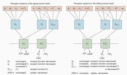
FIGURE 10.10. Receptor systems and their signal transduction mechanisms in the aging (left) and failing (right) human heart. β1, β2, α1 Stands for β1-, β2-, and α1-adrenoceptors, respectively; H2, histamine H2-receptors; 5-HT4, serotonin 5-HT4-receptors; M2, muscarinic M2-receptors; A1, adenosine A1-receptors; ETA, endothelin ETA-receptors; AT1, angiotensin II AT1-receptors; Gs, stimulatory G-protein; Gi, inhibitory G-protein; Gq/11, the G-protein that couples ET-, AT-receptors and α1-adrenoceptors to phospholipase C (PLC); AC, adenyl cyclase; PIP2, phosphatidylinositol 4,5-bisphosphate; DAG, diacylglycerol; IP3, inositol trisphosphate; GRK, G-protein-coupled receptor kinase; uptake1, noradrenaline reuptake transporter (+, activation; -, inhibition). (From Brodde OE, Leineweber K. Autonomic receptor systems in the failing and aging human heart: similarities and differences. Eur J Pharmacol 2004;500:168.)


CHANGES IN DRUG PHARMACOKINETICS DUE TO CARDIOPULMONARY BYPASS

CPB may affect the pharmacokinetics of drugs in a variety of ways, including changes resulting from hemodilution, hypothermia, altered organ perfusion, acid-base status, drug sequestration into the lungs and CPB circuit, and altered metabolism and clearance due to development of a systemic inflammatory response syndrome (SIRS) (Fig. 10.13) (90,91,92,93,94).


Hemodilution

The CPB apparatus is primed with fluid—usually some combination of crystalloid and colloid. At the time of initiation of CPB, the addition of this fluid to the circulation has several effects:


  • An immediate reduction in concentrations of circulating proteins such as albumin and a1-acid glycoprotein (α1AGP). This has implications for protein binding of drugs resulting from alteration in the ratio of bound drug to free drug in the circulation (5,6,7,8,9,10,95,96,97,98,99,100,101,102,103).


  • An immediate reduction in red blood cell concentration, which has implications for compounds that are sequestered to a significant degree in red blood cells (3,8,104,105).


  • An immediate reduction in the amount of free drug in the circulation at the initiation of CPB. This will reduce the amount of drug available for interaction with the receptor, with the potential for adverse events, for example, lightening of the level of anesthesia (5,6,7,36,106).


  • Alteration in organ blood flow, which may affect drug distribution and clearance (107).

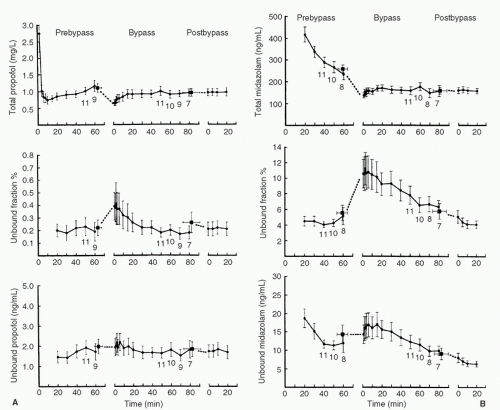
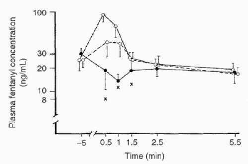
A number of studies have examined these issues and determined their relative importance to clinical practice (5,6,7,8,9,95,97,98,99,101,108). Typical findings include a reduction in total drug concentration in plasma, with increased free drug concentration, while on CPB (Fig. 10.14) (8,101). Although the clinical significance of this finding is unclear, others have described a transient (usually <5 minutes) reduction in both free and total drug concentration at the initiation of CPB as a result of hemodilution (Fig. 10.15) (3,36).

The explanation for free drug concentrations being sustained during CPB is a pharmacokinetic one. The large volume of distribution for most anesthetic agents is largely due to their high
lipid solubility relative to the volume of the CPB prime. Thus, following intravenous administration, the tissues serve as a reservoir that sequesters the drug. At the onset of CPB, when plasma concentrations fall due to hemodilution, the drug moves down its concentration gradient from the tissue stores to plasma. As protein concentrations fall, the free drug concentration increases to reestablish an equilibrium based on its solubility in plasma (8,11,101). Because free drug concentration is responsible for drug effect (109,110), the increase in free drug concentration from altered protein binding may explain the increased pharmacodynamic effect observed during and after CPB (9,10,111).


FIGURE 10.11. Agonist activation and coupling/signaling properties of β-adrenergic receptor subtypes. GRK, G-protein-coupled receptor kinase; βArr, β-arrestin; PDE, phosphodiesterase; PI3K, phosphatidylinositol 3-kinase; AC, adenyl cyclase; Gs, stimulatory G-protein; Gi, inhibitory G-protein; cAMP, cyclic adenosine monophosphate; PKA, protein kinase A; NOS, nitric oxide synthase; ERK, extracellular signal-regulated kinases. (From Lohse MJ, Engelhardt S, Eschenhagen T. What is the role of β-adrenergic signaling in heart failure. Circ Res 2003;93:897.)


Hypothermia

CPB is frequently conducted under varying degrees of hypothermia. The independent effect of hypothermic CPB on physiologic function has been extensively examined, and the findings of these studies have frequently been generalized to other patient populations (112). Although the mechanism is debated, hypothermia has anesthetic properties (113,114,115). Decreased body temperature appears to shift fluid from the intravascular to the interstitial space (116). This may alter the volume of distribution by shifting protein-poor fluid from the intravascular to the interstitial fluid compartment. Moreover, hypothermia activates autonomic and endocrine reflexes (117), which may produce peripheral vasoconstriction and alter the distribution of blood flow (118). Finally, hypothermia depresses metabolism by inhibiting enzyme function. As a consequence of these changes, pharmacokinetics may be altered through the following mechanisms:


  • Peripheral vasoconstriction may decrease absorption of drugs administered other than by the intravenous route (119).


  • Fluid extravasation may alter drug distribution from central to peripheral compartments (i.e., changes in the volume of distribution, Vd) (94,120).


  • Vasoconstriction may reduce the rate of reuptake of drug from peripheral tissues to the central compartment (120,121).


  • Temperature-induced reductions in enzyme-mediated biotransformation may decrease clearance of drugs and increase their elimination half-time (Fig. 10.16) (120,121,122,123,124,125,126,127,128,129,130,131).


  • Changes in organ perfusion may produce altered renal drug excretion as a result of decreased renal perfusion, glomerular filtration rate, and tubular secretion (132). However, studies comparing renal function during normothermic versus hypothermic bypass have not demonstrated any clinically important differences (117,130,133,134).


  • Hypothermia-induced increases in drug solubility in blood of volatile anesthetics (135).


FIGURE 10.12. Calcium cycling in cardiac myocytes and regulation by protein kinase A (PKA). AC, adenyl cyclase; RyR, ryanodine receptor; PLB, phospholamban; SERCA, sarcoplasmic reticulum calcium ATPase; CaM, calmodulin; CaMK, calmodulin-dependent kinase; CaN, calcineurin; GRK, G-protein-coupled receptor kinase; NCX, sodium-calcium exchanger; NHE, sodium-proton exchanger; PP, protein phosphatase; Gs, stimulatory G-protein; BAR, β-adrenergic receptor; PPI-1, protein phosphatase inhibitor-1; MyBPC-C, myosin binding protein C, slow type; P, phosphorylation. (From Lohse MJ, Engelhardt S, Eschenhagen T. What is the role of β-adrenergic signaling in heart failure. Circ Res 2003;93:897.)


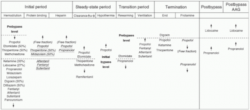
FIGURE 10.13. Plasma drug levels and factors affecting their concentration during cardiopulmonary bypass. All drug names indicate the level of plasma concentration unless the column denotes plasma free fraction change (italics and underlines). AAG increases after surgery. AAG, α1-acid glycoprotein. (From Mets B. The pharmacokinetics of anesthetic drugs and adjuvants during cardiopulmonary bypass. Acta Anaesthesiol Scand 2000;44:264.)


FIGURE 10.14. A: Total plasma concentration, unbound fraction, and unbound plasma concentration for propofol as a function of time for prebypass, bypass, and post-bypass periods. Each data point represents n = 12 (mean ± SEM). Numbers adjacent to the data points indicate n, where n is less than 12. The square data point to the left of t = 0 of the bypass period in each graph represents the mean of the final prebypass samples (mean ± SEM) plotted at the time (mean ± SEM) they occurred. The square data point to the left of the t = 0 of the post-bypass period in each graph represents the mean of the final bypass samples (mean ± SEM) plotted at the time (mean ± SEM) they occurred. Total plasma concentrations fall at the initiation of CPB, with little change in free drug concentrations, leading to an increase in the free fraction. B: Total plasma concentration, unbound fraction, and unbound plasma concentration for midazolam as a function of time for prebypass, bypass, and post-bypass periods. Each data point represents n = 12 (mean ± SEM). Numbers adjacent to the data points indicate n, where n is less than 12. The square data point to the left of the E = 0 of the bypass period in each graph represents the mean of the final prebypass samples (mean ± SEM) plotted at the time (mean ± SEM) they occurred. The square data point to the left of t = 0 of the post-bypass time period in each graph represents the mean of the final bypass samples (mean ± SEM) plotted at the time (mean ± SEM) they occurred. Total plasma concentrations fall at initiation of CPB, with little change in free drug concentrations, leading to an increase in free fraction. (From Dawson PJ, Bjorksten AR, Blake DW, et al. The effects of cardiopulmonary bypass on total and unbound plasma concentrations of propofol and midazolam. J Cardiothorac Vasc Anesth 1997;11:559.)

Hypothermia during CPB alters the clearance of drugs that require enzymatic degradation to terminate their effect (e.g.,
esmolol, remifentanil, clevidipine, atracurium, and cis-atracurium) (123,124,125,127). The net result is a prolonged drug effect that requires dosage reductions.


FIGURE 10.15. Plasma fentanyl concentrations (mean ± SD) in patients connected to cardiopulmonary bypass (CPB) circuits with primes containing no fentanyl (•—•) or containing a calculated fentanyl concentration of 140 (°—°) or 280 (°——°) ng/mL. X, lowest drug concentration measured at each stage during the first 1.5 minutes of CPB in patients not receiving fentanyl in their prime. NB: Regardless of whether the prime is supplemented or not, no difference exists in fentanyl concentrations within 2.5 minutes. (From Hynynen M. Binding of fentanyl and alfentanil to the extracorporeal circuit. Acta Anaesthesiol Scand 1987;31:708.)


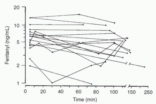
FIGURE 10.16. Fentanyl plasma concentrations in 18 children during profound hypothermia (18°C-25°C). Time zero is the initiation of cardiopulmonary bypass. Total plasma fentanyl levels remain essentially unchanged. (From Koren G, Barker C, Goresky G, et al. The influence of hypothermia on the disposition of fentanyl—human and animal studies. Eur J Clin Pharmacol 1987;32:374.)

For drugs with a low Vd, the vasoconstriction produced by hypothermia may even further decrease their Vd. This may explain the increase in plasma concentrations of neuromuscular relaxants observed during hypothermic CPB (94,136,137). When normothermia is being reestablished, reperfusion of tissues leads to washout of drug sequestered in underperfused tissues during the hypothermic CPB period. This may explain increases in plasma opioid concentrations observed during the rewarming phase (138) and postoperatively (139).


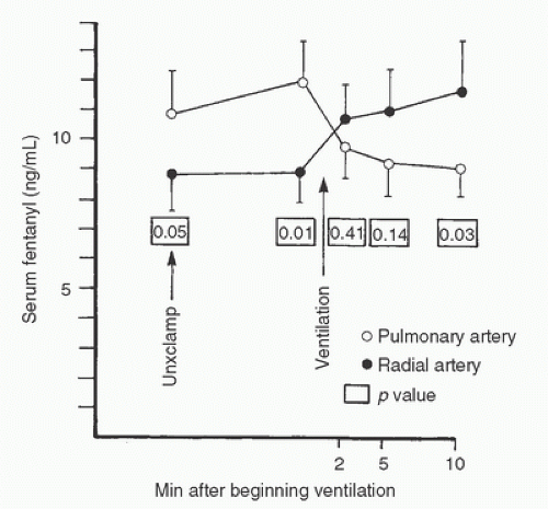
FIGURE 10.17. Mean fentanyl levels in seven cardiac surgery patients as ventilation and perfusion to the lung are resumed near the end of cardiopulmonary bypass. Systemic fentanyl concentrations rise with ventilation, whereas levels in the pulmonary artery fall, suggesting washout of fentanyl sequestered in the lungs during CPB. Unclamp, removal of aortic crossclamp. (Bently JB, Canahan TJ III, Cork RC. Fentanyl sequestration in lungs during cardiopulmonary bypass. Clin Pharmacol Ther 1983;34:705.)


Perfusion

CPB may be conducted with or without pulsatile perfusion (140). Nonpulsatile perfusion alters tissue perfusion (140). However, no difference in thiopental concentrations during CPB was detected when pulsatile versus nonpulsatile flow was studied (141). In contrast, as compared to those receiving nonpulsatile perfusion, cefamandole tissue concentrations were higher and elimination half-time prolonged in patients undergoing pulsatile perfusion (142). The degree to which pulsatile perfusion alters drug pharmacokinetics is therefore unpredictable and requires further study.

During CPB, the lungs are excluded from the circulation. Drugs taken up by the lungs (e.g., opioids (143,144,145,146,147), propofol (148), diazepam (149)) are therefore sequestered during CPB. Thus, the lungs may serve as a reservoir for drug release when normal circulation is reestablished (Fig. 10.17) (143). However, this effect is quite transient (143,146,147). In an animal model of drug administration post-CPB, regional concentration differences of the antibiotic levofloxacin (higher in upper lobes) persisted in lung fields which were not seen in an off-pump coronary artery bypass (OPCAB) (not receiving CPB) group suggesting that altered lung perfusion may persist post-CPB (150). Similar findings were demonstrated in cardiac surgery patients,
in whom postoperative lung concentrations of levofloxacin administered in the intensive care unit (ICU) were lower among those undergoing CPB versus a group not undergoing CPB. This finding was attributed to a higher degree of atelectasis (and hence altered tissue distribution) in postoperative CPB patients (151).

Although not a universal finding (152), most studies indicate that splanchnic blood flow is altered during CPB (19,118,122,153,154,155,156,157,158) and by drugs administered during CPB, such as vasopressin, dopamine, dobutamine, nitroglycerin (53,159), and anesthetic agents (160). This may affect the metabolism of drugs with a high hepatic intrinsic clearance (122) (e.g., fentanyl (56,90,120,161) and propofol (8,17,162)).


Acid-Base Status

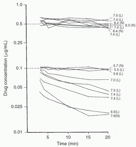
CPB may be conducted using pH-stat or a-stat blood gas management (163,164). While there is debate about which blood gas management strategy is best (165,166), the change in pH (164,167) with either of these schemes may affect organ blood flow (e.g., increased cerebral blood flow with pH-stat (168)), which may, in turn, affect drug distribution (163,168,169). pH management may also affect the degree of ionization and protein binding of certain drugs, leading to either increased or decreased free (active) drug concentrations (Fig. 10.18) (169,170).


Sequestration

Drugs may be taken up by various components of the CPB circuit itself (94). Various oxygenators have been reported to bind drugs in vitro, including volatile anesthetic agents (171,172,173,174,175), propofol (176,177), opioids (3,169,178,179,180), barbiturates (141), nitroglycerin (181,182), benzodiazepines (183), nifedipine (184), and antibiotics (91,185). For intravenous drugs, this phenomenon has rarely been demonstrated to be clinically important in vivo, likely because, depending on protein binding and lipophilicity, any free drug given intravenously and removed by the circuit is replaced from the much larger tissue reservoir (3,94,141,176). Nevertheless, unless the priming solution is primed with drug, and particularly for more hydrophilic drugs (e.g., antibiotics (186)), the potential exists for sequestration to lower the concentration below a minimum acceptable therapeutic level when CPB is initiated (94,185). This effect may be transient and counterbalanced by increases in plasma-free drug concentrations induced by reduced protein binding.

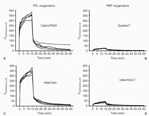
In the case of volatile agents added to the CPB circuit, wash in, wash out, and equilibration are accomplished quickly (104,105,187). Uptake by the oxygenator may occur in vitro (94,179), but in vivo uptake typically is clinically insignificant. Exhaust gas measurements from the oxygenator have been used as surrogate measures to indicate arterial concentrations of volatile agents. However, the anesthetic levels reported by Wiesenack et al. (173) have drawn our attention to the possibility of difficulties with the plasma-tight poly-(4-methyl-1-pentene) (PMP) type of oxygenator. Following introduction of these oxygenators into their practice, they noted an increased incidence of elevated perfusion pressure (indicative of light anesthesia) in patients undergoing CPB who were receiving a volatile agent (13). They examined this issue by performing an in vivo comparison of the uptake of isoflurane by two types of microporous polypropylene (PPL) oxygenators versus two types of PMP oxygenators. Significant differences in gas transfer rates were observed between the two groups (Fig. 10.19) (173). They attributed this difference to a significantly reduced diffusion coefficient for the volatile agent in the solid layer of the new membrane. Similar findings were reported by Prasser et al. (175). Hinz et al. reported that the exhaust gas measurement of sevoflurane showed washout of the gas in the PPL group during CPB but not in the PMP group. However, while they declined in the PLP group, plasma concentrations of sevoflurane in this study were steady in the PMP group during CPB in the face of a constant gas concentration and flow rate. The decline in sevoflurane concentration in the
PLP group was associated with an increase in blood pressure and Bispectral Index (BIS) scores (BIS—a measure of anesthetic level), indicative of lightening of anesthesia, a finding which was not observed in the PMP group.


FIGURE 10.18. Changes in the concentrations of alfentanil (top) and fentanyl (bottom) in extracorporeal circuit prime with time (shown on the same logarithmic scale—vertical axis). Dotted lines represent predicted concentrations. Numbers on the right side are pH values of each priming solution. L, low temperature (24.4°C-25.70°C); N, normothermic (34.1°C-37.0°C); NB: The differences in binding to the cardiopulmonary bypass apparatus occur as a result of dissimilarities in the ionization of the two drugs with changes in pH. (From Skacel M, Knott C, Reynolds F, et al. Extracorporeal circuit sequestration of fentanyl and alfentanil. Br J Anaesth 1986;58:948.)


FIGURE 10.19. Isoflurane blood concentrations (Cisoflurane [µm]) for the uptake and elimination sequence in the four oxygenator groups. Each line represents a single patient. During hypothermic cardiopulmonary bypass, isoflurane 1% was administered to each patient. A: CapioxRX25; B: Hilite7000; C: QuadroxD; D: Hilite7000LT. (From Wiesenack C, et al. In vivo uptake and elimination of Isoflurane by different membrane oxygenators during cardiopulmonary bypass. Anesthesiology 2002;97:133-138.)

The above results suggest that the use of exhaust gas measurement of volatile anesthetic concentrations as surrogates for arterial concentrations or anesthetic effect may be inappropriate in some circumstances. Thus, as newer technology becomes available, each membrane must be tested to determine its relative ability to transport volatile agents during CPB in order to prevent inadequate levels of anesthesia at the time of separation from CPB.

The red blood cell may serve as a reservoir for drugs, and the anemia associated with CPB may alter drug concentrations (3). The clinical relevance of this mechanism remains uncertain. Drug concentrations in red blood cells increase during CPB—presumably a reflection of the increased distribution of drugs as a result of hemodilution (8).

Hemofiltration is often performed to remove excess fluid administered during CPB, particularly in pediatric cardiac surgery and in patients with renal dysfunction (188,189,190,191). Factors affecting the degree to which a drug is removed by hemofiltration are listed in Table 10.3 (192). Koster et al. (190) examined the ability of four hemofilters (Renoflow 11, Baxter; Arylane H4, Cobe; Ultraflux AV 600, Fresenius; and BCS 110 Plus, Ios-tra) and two plasmapheresis filters (ASAHI Plasmaflow OP, Diamed; PF 2000 N, Gambro) to remove hirudin in an in vitro simulation of CPB. They determined that the plasmapheresis filters were more efficient at removing hirudin. O’Rullian et al. (193) determined that cefazolin concentrations were not different when a hemofilter was employed as part of the circuit. Similarly, aprotinin concentrations were not reduced by hemofiltration (194). In contrast, tirofiban was removed to the same degree in an in vitro study comparing two hemofilters (Hospal Arylane H4 and Minntech Hemocor HPH 700) and a plasmapheresis filter (ASAHI Plasmaflow OP) (195). In a pediatric population, use of modified ultrafiltration reduced the concentration of the poorly protein-bound antibiotic flomoxef by an amount equivalent to that removed by the kidney (196). It therefore appears that the degree to which drugs are removed by hemofiltration during CPB will require further study of individual drugs and filters. While in
theory the use of a cell saver device might have an effect on clearance of protein-bound drugs, this has not been demonstrated to be a clinical concern to date (197).


TABLE 10.3. Factors influencing movement across a membrane


Protein binding of the solute


Volume of distribution (Vd)


Solute/membrane interaction


Solute charge


Protein concentration (which influences both solute binding and oncotic pressure)


Ultrafiltrate line suction


Blood flow through hemofilter


Viscosity of blood (degree of hypothermia)


Length and diameter of tubing


Venous resistance


Filter surface area and properties


Reprinted from Clar A, et al. Derivation of sieving coefficients to determine the efficacy of the hemoconcentrator in removal of four inflammatory mediators produced during cardiopulmonary bypass. ASAIO J 1997;43:63-170.


CHANGES IN PHARMACODYNAMICS DURING CARDIOPULMONARY BYPASS

The ability of a drug to produce its effect depends on the ability of free (unbound) drug to reach its receptor, bind with it, and translate the receptor-initiated signal to its effectors (109,110,198). A number of factors encountered during cardiac surgery and CPB may affect this sequence of events.


Protein Binding

In the blood, drugs exist as free (unbound) drug in equilibrium with the bound (i.e., bound to plasma proteins) drug. Only free drug is capable of interacting with its receptor and producing pharmacologic effects (9,110). In plasma, drugs bind primarily to the proteins albumin (acidic drugs) and a1AGP (basic drugs, e.g., fentanyl, lidocaine), an acute-phase reactant (110). The concentration of a1AGP rises during stress, including that produced by the systemic inflammatory response initiated during CPB, and this reduces lidocaine (199,200,201), quinidine (201), and propranolol (201) free drug concentrations following CPB. This may result in the return of arrhythmias despite adequate measured total drug concentrations after CPB.

Changes in protein binding are clinically significant only for drugs that are highly protein-bound (11). The degree of drug-protein binding depends on the total drug concentration, the available protein concentration, the affinity of the protein for the drug, and the presence of other substances that may compete with the drug or alter drug binding sites (202). Measurement of total drug concentrations in plasma during CPB may therefore fail to elucidate the true picture of changes in drug effect unless the unbound concentration is also measured (Fig. 10.14) (6,8,11,101).

The degree of protein binding may be altered by pathologic states existing before CPB, for example, acute myocardial infarction (MI) (200), renal disease (203), and diabetes (204), or as a result of changes occurring during CPB, for example, development of the SIRS, leading to a reduction in albumin production and an increase in a1AGP concentrations (93,108,199,201,205). In the presence of renal failure and liver disease there may be reduced plasma albumin concentrations (206). Further, the affinity of albumin for drugs such as phenytoin, thiopentone, and diazepam may be reduced in the presence of chronic disease states (e.g., cirrhosis) (15,206,207).

Heparin is administered before and during CPB to prevent clot formation. Heparin releases free fatty acids, which may in turn displace drugs from protein binding sites and increase free drug concentrations, resulting in an enhanced pharmacologic effect (96,208,209,210).


Tissue Binding

Following drug administration, free drug penetrates into tissues where it may bind to tissue proteins. Certain tissues, for example, those of the heart and lung, have an affinity for certain drugs (29,143,144,145,146,147,148,209,211,212). This has two relevant pharmacodynamic consequences. First, tissue binding may limit access of a drug to its receptor if that tissue is not the site of the desired drug action, thereby potentially limiting the magnitude of effect (147). Second, the tissue may also serve as a drug reservoir, thereby extending drug effect, particularly if the tissue (e.g., lung) is out of circuit during CPB (Fig. 10.17) (143).


Age

Adult cardiac surgical patients are frequently elderly (defined as an age > 65 years). Independent of pathologic processes, aging is associated with a variety of physiologic processes that may influence drug effect (15). The functions of organs such as the heart are reduced while blood flow to the kidneys and liver are decreased (213). These factors may reduce drug clearance (214). Elderly humans also have increased adipose tissue, which may serve as a reservoir for lipid-soluble drugs. Albumin concentrations may be reduced, resulting in higher free drug concentrations when standard doses are administered (15,214).

A number of investigations have recently examined the sensitivity of the central nervous system (CNS) to drugs in the elderly. While CNS sensitivity to barbiturates may be unaltered (215), reductions in dose are required because of a
slower return of drug from the effect site to the central compartment (a pharmacokinetic difference) (216). In contrast, a true difference in CNS sensitivity to opioids (fentanyl, alfentanil, and remifentanil) (217,218) and volatile agents (219) has been described in the elderly. For the elderly patient undergoing CPB, a reduction in dose to limit the high free drug concentrations and the potential for prolonged or toxic drug effects therefore seems prudent.

At the other end of the age spectrum, infants and children may have altered drug effects owing to differences in distribution of body fat, maturity of clearance mechanisms, and disease-related changes in drug handling (38,220,221,222,223,224,225,226,227,228,229). Although the degree to which infants and children may have differences in drug sensitivity has received little scientific scrutiny, differences appear to exist. Thus, it cannot be assumed that doses of drugs considered adequate for adults undergoing CPB will suffice for the pediatric population (222,224,230,231).


Central Nervous System Penetration

For anesthetic agents, the site of drug effect is presumed to be the CNS. Differences in CNS pharmacodynamic effects between drugs have been measured by comparing the time difference between their peak plasma concentrations and electroencephalographic (EEG) changes (232). A hysteresis (i.e., less time to equilibrium with alfentanil) has been observed between the peak plasma concentration of fentanyl versus alfentanil and the shift in the spectral edge frequency of the EEG (233). A similar time lag has been observed as drug concentrations fall. These findings most likely represent disposition of the more lipophilic fentanyl to highly lipophilic brain tissue, preventing fentanyl concentrations from rising at the CNS opioid receptors and prolonging time to peak effect (a pharmacokinetic difference). This hypothesis is substantiated by the demonstration that hysteresis for sufentanil (another lipophilic opioid) more closely approximates that of fentanyl than the more hydrophilic alfentanil (234). A similar rationale has been used to describe differences in onset of action between midazolam and diazepam (232,235). Utilization of these time differences and concentrations can be measured and modeled, and has led to the development of computer programs with a rate constant (Ke0) designed to target stable drug concentrations at the effect site compartment (receptor level) (31,37,236). Such programs have been utilized in studies of drug administration to target concentrations during cardiac surgery (28,37,40,41,42,224). By providing stable plasma concentrations, determination of concentration-versus-effect relations during CPB can be made by utilizing various monitors of CNS activity, such as auditory evoked potentials (AEP) (237) and the BIS (10,21,25,28,31,32,33,34,35,39,40,42,114,238,239,240).

The lysine analogs e-aminocaproic acid and tranexamic acid are commonly employed to prevent excessive bleeding during cardiac surgery. Although these agents effectively inhibit the fibrinolytic system that is activated during CPB (241), their use has unfortunately been associated with development of seizures in the postoperative period (242). In part, this may be attributable to the variable penetration of these drugs into the CNS. In a study of tranexamic acid, cerebrospinal fluid (CSF) levels of the drug differed 9-fold despite administration of a fixed body weight dose (243). At clinically relevant CSF concentrations, the mechanism for production of seizures with tranexamic acid may be related to blockade of the receptor for the inhibitory neurotransmitter glycine, which has been prevented in animal models by administration of propofol or isoflurane (244).


Temperature

Hypothermia produces a number of pharmacodynamic effects. Anesthetic requirements are reduced by hypothermia (113,114,245). Changes in receptor affinity (e.g., decreased opioid receptor affinity (246) and nicotinic acetylcholine receptor sensitivity (247)) also occur during hypothermia. The action of neuromuscular receptor blocking agents is enhanced, which may reflect a pharmacodynamic and a pharmacokinetic effect (91,130,137,248,249,250,251,252) (Table 10.4).

Use of hypothermia may decrease release of the excitatory neurotransmitters glutamate and glycine into the CNS (253), thereby attenuating central excitotoxicity and exerting a cerebroprotective effect. The implications of such an effect on drug action are unknown, but hypothermia has not reduced the incidence of neurocognitive deficits in most (but not all (254)) prospective randomized clinical trials (238,255).


TABLE 10.4. Muscle relaxants and hypothermic cardiopulmonary bypass


Plasma concentration on CPB


Medication


Initial


Later


Clearance


N-M sensitivity


Steroids


Pancuronium


↑ 1.8-fold


Vecuronium


↑ 5-fold


Rocuronium


Benzylisoquinoliniums


d-Tubocurarine


Metocurine


Atracurium


cis-Atracurium


↑ 2-fold


Toxiferines


Alcuronium


N-M, neuromuscular; ↑, Increase; ↓, decrease; •, not known.


Modified from Mets B. The pharmacokinetics of anesthetic drugs and adjuvants during cardiopulmonary bypass. Acta Anaesthesiol Scand 2000;44:261-273.


Acid-Base and Electrolyte Changes

Altered peripheral perfusion produces tissue acidosis during CPB (256). This may affect the response to catecholamines (257). The degree of ionization and protein binding (hence free drug concentrations) of weak acids and bases may also be affected by the blood gas management strategy employed during CPB (Fig. 10.18) (169). Changes in pH may also affect electrolyte balance. Calcium, magnesium, and potassium concentrations decline during CPB (258,259,260), and associated muscle weakness, arrhythmias, and enhanced digitalis toxicity may ensue. Whether intraoperative replacement of calcium (261,262) or magnesium (263,264) reduces complications such as arrhythmias is still debated.


Cardiopulmonary Bypass

Volatile anesthetic agents are commonly employed during CPB, usually in combination with intravenous anesthetic agents to avoid hemodynamic aberrations (265,266). Volatile agents may disrupt ion channels and intracellular transduction mechanisms (267). Although the nature of the priming solution (acetate) may prove important by acting as a supplemental anesthetic agent, the anesthetic requirements for volatile agents are reduced by an as of yet unidentified mechanism following CPB in dogs (268,269,270).

Using the brain’s electrical activity and “depth of anesthesia” monitors (which may be problematic in terms of sensitivity (9,114,239)) to examine the independent role of CPB in reducing anesthetic requirements in man has yielded mixed results. Yoshitani et al. (10) demonstrated a greater pharmacodynamic effect for a given concentration of propofol pre- versus post-CPB under normothermic conditions. Takizawa et al. (9) similarly reported an enhanced propofol effect for a given dose following normothermic CPB, which was in part related to an increased free (but not total) drug concentration during CPB. In contrast, Ahonen et al. (271) examined the need for propofol to maintain a fixed BIS in a group of patients randomized to on- versus off-pump surgery, and could not detect any difference in anesthetic requirements. A randomized trial of patients undergoing cardiac surgery by Mathew et al. (245) reported that CPB produced a reduction in BIS levels that was further enhanced with use of hypothermia. Lundell et al. (272) examined the concentration-effect relation for isoflurane in combination with a computer-driven infusion of fentanyl designed to maintain a constant effect site concentration of 3 ng/mL and noted a 25% reduction in the concentration of isoflurane required to maintain a stable BIS level post- versus pre-CPB. These findings suggest that the CPB itself may reduce anesthetic requirements.


Receptor Density

The number of receptors available for interaction with a ligand will determine the magnitude of drug effect. Patients presenting for cardiac surgery frequently have CHF (266,273). These patients frequently have a reduced number of cardiac β-receptors, defects in receptor transduction, and impaired synthesis and reuptake of norepinephrine (78,88,274). Chronic β-adrenergic receptor agonist administration has a pharmacodynamic effect (275), and continued use of these agonists has been linked to therapeutic failure, including increased mortality (276). As chronic administration of β-adrenergic receptor blocking agents in those with CHF improves morbidity (277) and mortality (276,277,278), affected patients typically take these agents preoperatively. As a consequence of both a reduced number of receptors and pharmacologic blockade of β receptors, β-adrenergic agonists may exhibit reduced pharmacodynamic effects during and following CPB (279).

Discontinuation of β-adrenergic receptor blocking agents in patients treated for angina pectoris and CHF in the perioperative period may lead to β-adrenergic receptor upregulation and increased adrenergic responsiveness (280). Increases in myocardial oxygen demand due to increased heart rate and contractility may lead to myocardial ischemia, worsened symptomatology, and even death (281). A retrospective database analysis suggested that continuation of β-adrenergic receptor blocking agents might also have a neuroprotective effect (282). It is therefore generally recommended that chronic cardiovascular medications, including β-adrenoceptor receptor blocking agents, be continued until surgery commences (280).

Changes in receptor density and function may occur quickly (77), even during the conduct of cardiac surgery (283). Changes in the density and function of the β-adrenergic receptor have been examined the most among the cardiac surgery population. β-adrenergic receptor function is impaired following CPB (284). Possible mechanisms include ischemia-reperfusion injury (285) and acute β-adrenergic receptor desensitization (286) occurring as a result of the catecholamine release accompanying CPB (117). At the cellular level, there is a reduction in GRK activity (74). Attempts to ameliorate β-adrenergic receptor dysfunction during CPB have included administration of β-adrenergic receptor blocking agents (287) and the use of warm blood cardioplegia (288) or high spinal anesthesia (289). Further work is required to elucidate the mechanisms and identify the best therapeutic options to reduce undesirable changes in receptor density and function.


Hyperalgesia

Tissue injury, including surgical trauma, may result in hyperalgesia (exaggerated nociceptive responses to noxious stimuli) and allodynia (nociceptive responses to innocuous stimuli) (290,291). Although the mechanism(s) by which this occurs is likely multifactorial (292), emerging evidence suggests that excitatory amino acids (e.g., N-methyl-D-aspartate (NMDA) at the spinal cord level may play a key role (290). Stimulation
of excitatory amino acid receptors in the spinal cord triggers intracellular events leading to Ca2+ release and phosphokinase C (PKC) activation. PKC is associated with a number of neuronal changes, including development of hyperalgesia (290). As tolerance to morphine administration shares many of the same pathways (290), studies in animals and humans have also implicated these pathways in the development of hyperalgesia (so-called opioid-induced hyperalgesia [OIH]) (291,293). Whether hyperalgesia develops in the perioperative period is debated, but recent meta-analysis concluded that, at least for the administration of remifentanil, it does (294). Representative studies, in patients undergoing cardiac surgery, include that of Richebe and colleagues (295) who examined the incidence of hyperalgesia following remifentanil administration by CACI with a target concentration of 7 ng/mL as compared to a group of patients receiving a zero-order infusion of 0.3 µg/kg/min. Use of the targeted infusion led to a reduction in total remifentanil dose and a reduction in hyperalgesia. In a randomized study of 40 patients undergoing coronary artery bypass graft (CABG) surgery comparing sufentanil targeted to a plasma concentration of 0.4 or 0.8 ng/mL, it was observed that the higher-dose sufentanil group had increased postoperative analgesic requirements, but assessments for hyperalgesia indicated that, while present and diminishing over time, the degree of hyperalgesia was not different between the two groups (296). In contrast, in a randomized study of 90 patients undergoing cardiac surgery and receiving either sufentanil 0.3 µg/kg/min intraoperatively or placebo (in addition to a sufentanil/propofol-based anesthetic), no difference in postoperative analgesic requirements was observed. Although the authors concluded that hyperalgesia did not occur (297), it must be acknowledged that it was not specifically tested for as in the studies of Fechner et al. (296) and Richebe et al. (295).

Therapeutic approaches to the management of OIH have varied (298). The role of ketamine (an NMDA receptor antagonist) was examined in a group of patients undergoing abdominal surgery and receiving high doses of remifentanil intraoperatively (299). Ketamine administration reduced the degree of hyperalgesia.

The results of these investigations suggest that use of opioids should be restricted to what is necessary to control pain and unwanted hemodynamic responses so as to minimize the development of hyperalgesia in the perioperative period. Measures such as administration of ketamine to reduce hyperalgesia when recognized merit further investigation.


THE SYSTEMIC INFLAMMATORY RESPONSE SYNDROME

Cardiac surgery and CPB initiate a systemic inflammatory response as a consequence of operative trauma, blood contact with foreign surfaces with resultant complement activation, development of ischemia-reperfusion injury, and the presence of endotoxin (Fig. 10.20) (205). The magnitude of the response is influenced by a number of factors (93,205,300,301), including genetics (302), and may have an adverse effect on clinical outcomes (92,303,304), including development of multiple organ dysfunction syndrome (MODS) (19,134,154,305,306). The pathophysiologic mechanisms by which this occurs are gradually being elucidated (307,308,309,310,311,312,313).

Attempts to ameliorate the systemic inflammatory response are of interest as inflammation may have a profound effect on drug handling (Fig. 10.20) (92,314). During evolution of the inflammatory response, a host of cytokines and other mediators are released, including tumor necrosis factor alpha (TNF-α), interleukin 1 (IL-1), IL-6, IL-2, IL-8, IL-10, interferon γ (IFN-γ) and nitric oxide (NO) (93,205). Each of these mediators (e.g., TNF-α, IL-1, and IL-6 (93,315,316)) subserves a number of functions, including increasing transcription of acute-phase proteins (which may reduce free drug concentrations (108,199,201)) and inhibition of drug metabolism through downregulation of cytochrome P-450 activity (162,317). The metabolism of most clinically important drugs, including those utilized for the provision of cardiac anesthesia, is restricted to a relatively small number of cytochrome P-450 (CYP-450) isoenzymes, including CYP 1A2, 2A6, 2C8, 2C9, 2D6, and 3A4 (45,318,319,320). Of particular note, cytochrome P-450 3A4 (CYP3A4) is partly responsible for the metabolism of many of the opioids (321), propofol (322), and is exclusively responsible for the metabolism of midazolam (323,324).

Factors that affect CYP3A4 function can thus have a significant effect on drug metabolism. In addition to inflammatory cytokines (325), these include endotoxin (326), drugs (327,328,329,330), food (331), hypoxia (332), neurotransmitters (including adrenaline) (333), CPB (334), preexisting disease (335), and race (336). The variability in drug handling seen during cardiac surgery may be partly explained by these factors.

Myocardial depression may be induced by IL-6 (93,337,338) and NO (338), which may, in turn, alter the distribution of drugs. Activation of neutrophils with generation of oxygen-derived free radicals may injure tissue, particularly endothelium, leading to a capillary leak syndrome that may affect the volume of distribution for drugs and drug penetration into tissue receptor sites (339,340,341,342

Only gold members can continue reading. Log In or Register to continue

Stay updated, free articles. Join our Telegram channel

Jun 7, 2016 | Posted by in RESPIRATORY | Comments Off on Changes in the Pharmacokinetics and Pharmacodynamics of Drugs Administered during Cardiopulmonary Bypass

Full access? Get Clinical Tree
Get Clinical Tree app for offline access