Fig. 30.1
(a) Aortic root angiogram in anteroposterior view shows tortuous coronary artery fistula (CAF) (arrow) arising from the left coronary artery (LCA) entering right atrium (RA). Normal branching of the LCA into the left anterior descending (LAD) and left circumflex (Cx) is well seen. Proximal portion of the right coronary artery (RCA) is also opacified. (b) It was closed with two Gianturco coils (arrow)
Case Study 2
A 4-month-old asymptomatic infant was incidentally found to have a murmur. His echo revealed a CAF from RCA to RA. On follow-up, there was a progressive enlargement of the fistula from 5 mm at 4 months to 11 mm at 1 year of age. The rapid enlargement of the feeding artery on echocardiography indicated closure of fistula despite absence of symptoms (Fig. 30.2).
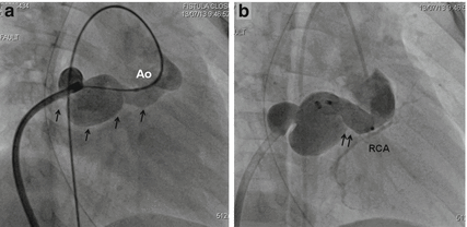

Fig. 30.2
(a) Angiogram through venous sheath after AV loop formation in right anterior oblique projection shows a large fistula (arrows) from the proximal part of right coronary artery (RCA) coursing posteriorly to enter the right atrium. Faint opacification of the aortic root (Ao) is also seen. The RCA branches are not well seen due to high flows through the fistula. (b) After closure with a 14–12 duct occluder I device (arrows), RCA fills well
Case Study 3
Angiogram of a 15-day-old presenting with heart failure showed a large fistula from LCA to the RA coursing posterior to the aortic root. Surgery was performed through midline sternotomy, and a large coronary fistula from the left coronary sinus behind the aortic root was identified in the transverse sinus of the heart. The fistula was clipped in its course without the use of cardiopulmonary bypass. Disappearance of the thrill was taken as confirmation of closure of the fistula and sternotomy was closed. The neonate remained ventilator dependent due to significant residual flow through the fistula. Lung infections complicated the course of the child further. Transcatheter closure of the fistula was prompted by persistent heart failure, growth failure, and recurrent pneumonia warranting ventilatory support (Fig. 30.3).
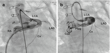

Fig. 30.3
(a) Angiogram through venous sheath after arteriovenous (A-V) loop formation in a patient with a significant residual flow from left coronary artery (LCA) to right atrial (RA) fistula after surgical ligation shows the long fistulous tract and normally branching LCA into the left anterior descending (LAD) and the left circumflex (Cx) arteries. Sternal wires and multiple clips placed to close the fistula surgically are seen. (b) Repeat angiogram after placement of Amplatzer vascular plug II through the venous sheath and an additional coil at the most distal end, shows a complete closure of the fistula with better visualization of the branches of the LCA
Case Study 4
A 7-year-old asymptomatic child was followed up for CAF from the RCA to the right ventricle (RV). Oximetry showed Qp/Qs of 1.7:1. Magnitude of the left-to-right shunt prompted transcatheter closure of the fistula (Fig. 30.4).
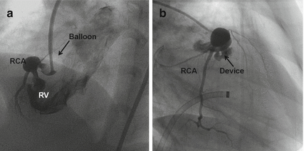

Fig. 30.4
(a) Aortogram in left anterior oblique view with cranial angulation using a balloon tipped catheter shows large fistula from proximal right coronary artery (RCA) to the right ventricle (RV). The branches of the RCA distal to the fistula are seen due to proximal balloon occlusion. (b) After closure with a duct occluder I, angiogram in right anterior oblique view shows the RCA with branches
Case Study 5
A 21-year-old asymptomatic man was identified to have a large fistula from the LCx to the ostium of coronary sinus on a preemployment medical examination. Angiogram showed a fistula arising from a markedly dilated LCx, measuring 22 mm, draining into the ostium of the coronary sinus, with a 1.9:1 shunt and mildly elevated pulmonary artery and LV filling pressures (Fig. 30.5).
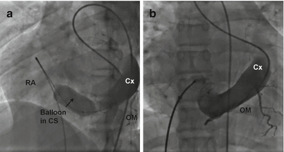

Fig. 30.5
(a) Selective angiogram in the left circumflex artery (Cx) after occluding the fistula opening into the right atrium (RA) by a 25 mm Tyshak II valvuloplasty balloon shows the obtuse marginal (OM) branches of the Cx. (b) After the distal end of fistula was closed with a large duct occluder I, the contrast is seen to opacify the Cx and OM branches more intensely
Case Study 6
An 8-year-old asymptomatic child with RCA to the RV fistula had entire RCA which was aneurysmally dilated to 12 mm from its aortic origin to the crux of the heart where the posterior descending interventricular branch (PDA) was given off. The fistula terminated immediately before the origin of the PDA. Even though the shunt was only 1.4:1, transcatheter closure was indicated by a threat of rupture of the aneurysmal RCA, which measured 12 mm (Fig. 30.6).
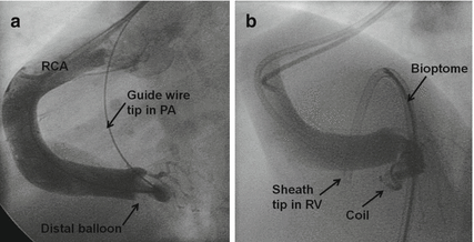

Fig. 30.6
(a) Selective right coronary artery (RCA) angiogram in left anterior oblique view shows a large RCA to right ventricle (RV) fistula. Through a second arterial access a guidewire was advanced through the fistula into the pulmonary artery and a distal balloon occlusion was done. The posterior descending and posterolateral branches of the RCA are better visualized only after the distal balloon occlusion. (b) The distal end of the fistula was closed from the venous side with bioptome assisted delivery of two intertwined embolization coils
Case Study 7
A 40-year-old man was diagnosed to have a small fistula from the atrial branch of the left circumflex artery to the right atrium during device closure of secundum atrial septal defect. After 6 years, he developed effort angina with reversible perfusion defect on myocardial perfusion sestamibi nuclear scan. The effort angina and nuclear perfusion defect were a result of myocardial steal through the fistula (Fig. 30.7).
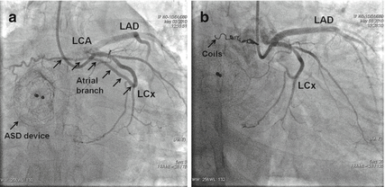

Fig. 30.7
(a) Left coronary artery (LCA) injection through right radial arterial access in right anterior oblique view shows a small fistula (multiple arrows) from the left circumflex artery (LCx) coursing posteriorly and terminating in the right atrium in a patient who had closure of atrial septal defect with an Amplatzer septal occluder (single arrow) earlier. The left anterior descending (LAD) artery is seen to be normal. (b) This fistula was closed with six micro coils (0.018” Hilal embolization coils, Cook medical) using a microcatheter through a left coronary artery guide catheter
Case Study 8
A 3-year-old asymptomatic young boy was diagnosed to have a fistula from a single left coronary artery to the right ventricle. The LAD continued beyond the apex in the posterior interventricular groove as the posterior descending artery (PDA) and subsequently coursed in the posterior right atrioventricular groove and terminated in a sac before entering the right ventricle. There was a large right-sided branch from the LAD that coursed in the right anterior interventricular groove which also terminated in the same sac before entering the right ventricle. The entire myocardial supply in the region of the right coronary artery in the right atrioventricular groove and the posterior interventricular groove was given off from the LAD (Fig. 30.8).
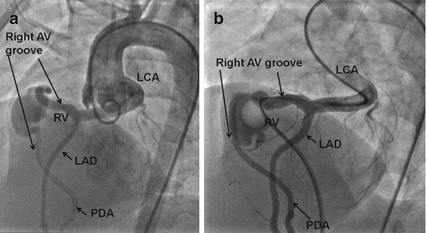

Fig. 30.8
(a) Aortic root angiogram in left anterior oblique projection shows absence of right coronary artery with faint opacification of the left coronary artery (LCA). A dilated left anterior descending (LAD) artery continues beyond the apex in the posterior interventricular groove as posterior descending artery (PDA) and then subsequently courses in the posterior right atrioventricular groove in region of RCA and drains finally into the right ventricle (RV). There is another anterior branch from the LAD that courses in the anterior right atrioventricular groove in the region of RCA and again enters into the fistulous sac. (b) In this high flow fistula, all the branches viz. left anterior descending (LAD), posterior descending (PDA) are better delineated by a selective LCA angiogram with distal occlusion done with a balloon wedge catheter from the right ventricle (RV)
30.4 Indications
1.
Heart failure and growth impairment
2.
Clinical features of large left-to-right shunt
3.
Enlarged heart on X-ray
4.


Echocardiographic evidence of dilated left ventricle and diastolic flow reversal in aorta
< div class='tao-gold-member'>
Only gold members can continue reading. Log In or Register to continue
Stay updated, free articles. Join our Telegram channel

Full access? Get Clinical Tree


