Case 7

Case 7


HISTORY AND PHYSICAL EXAMINATION


In a 72-year-old woman, pain developed in the right anterior thigh and knee, became severe over 2 to 3 weeks, and did not respond to steroid injection into the knee joint. The pain was maximal at night and did not worsen with standing or walking. Within 1 month, she noticed that her right leg was weak because the knee frequently would buckle from underneath her. She had fallen many times. She denied any symptoms in the left leg. She denied numbness in the legs or hands. She had mild low back pain. Her medical history was significant for diabetes mellitus, hypertension, and hypercholesterolemia. She was taking glyburide and diltiazem.


On physical examination, the patient was in modest discomfort because of right leg pain. Mental status and cranial nerve examinations were normal. Straight and reversed straight leg testing were negative bilaterally. Motor examination revealed mild atrophy of the right quadriceps muscle. There were no fasciculations. Muscle tone was normal. Manual muscle examination revealed severe weakness of right knee extension (Medical Research Council (MRC) 4–/5), thigh adduction (MRC 4–/5), and hip flexion (MRC 4/5). All other muscle groups were normal. Deep tendon reflexes revealed were normal in the upper extremities. In the lower extremities, the left knee jerk was normal, but the right knee jerk was absent and both ankle jerks were trace. Sensory examination was normal, both distally and in the anterior thighs. Gait was impaired by the right leg weakness. Romberg test was negative.


The patient was referred to the electromyography (EMG) laboratory.


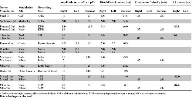
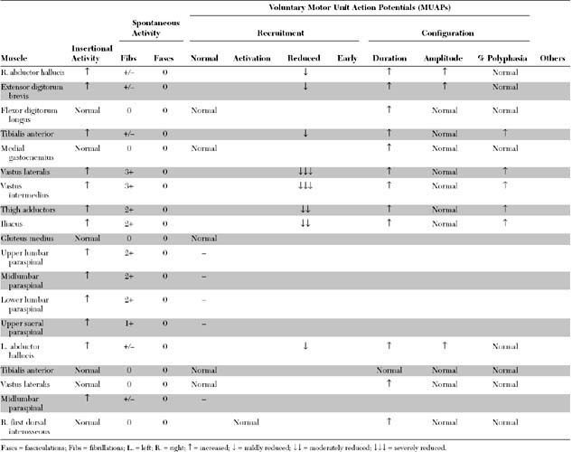
Please now review the Nerve Conduction Studies and Needle EMG tables.


EDX FINDINGS AND INTERPRETATION OF DATA


Relevant EDX findings in this case include:


These findings are consistent with a subacute severe right lumbar polyradiculopathy, affecting the L2–L4 roots, combined with mild chronic sensorimotor peripheral polyneuropathy. With unevokable saphenous SNAPs bilaterally in this patient with diabetes, the lesion could be anatomically a lumbar plexopathy, a lumbar radiculopathy, or more likely, a radiculoplexopathy. This case is compatible with the EMG findings seen in diabetic amyotrophy (diabetic proximal neuropathy). It is also common to find minor denervation in the contralateral limb, even when asymptomatic.


Discussion


Definition and Classification


Diabetes mellitus is a common disorder of two types. Type 1 is characterized by a severe or complete absence of insulin and is due to an autoimmune attack on the islets of Langerhans in the pancreas. Type 2 is more common accounting for about 90+ of diabetics in the United States, and is characterized by insulin resistance and influenced by many factors including obesity, diet, physical activity, and inheritance.


Diabetes mellitus has a propensity to cause microvascular disease, nephropathy, retinopathy, and peripheral neuropathy. Because of the heterogeneity of diabetic peripheral nervous complications, there is a lack of agreement among clinicians regarding the definition of diabetic neuropathy. An accepted definition of diabetic neuropathy is the presence of a clinical or subclinical diffuse disorder of somatic and/or autonomic parts of the peripheral nervous system in the setting of diabetes mellitus and in the absence of other causes of peripheral neuropathy.


The classification of diabetic neuropathies cannot be rigid because many overlap syndromes may be seen. A practical categorization, based on clinical presentation rather than precise etiology, divides these neuropathies into distal symmetrical polyneuropathy (the commonest), asymmetrical polyradiculoneuropathy, cranial mononeuropathy, and entrapment mononeuropathy (Table C7-1). Detailed discussions of all these syndromes are beyond the scope of this section and have been summarized in recent reviews (see Bird and Brown 2002; Brown and Asbury 1984; Harati 1987; Wilbourn 1993).


Table C7-1 Classification of Diabetic Neuropathy


* Common mononeuropathies with increased incidence in diabetics.


Clinical Features


Diabetic Sensorimotor Autonomic Polyneuropathy


Diabetic neuropathies are by far the most prevalent peripheral neuropathies encountered in clinical practice. Among all diabetic neuropathies, the mixed sensory-motor-autonomic peripheral polyneuropathy is by far the most common, and is usually related to the duration and severity of hyperglycemia. However, this form may occasionally be the presenting symptom of occult diabetes mellitus. The exact incidence of this diabetic polyneuropathy is not known; this is, in part, because of its diverse clinical presentations and different measurements used to define the presence or absence of neuropathy (Table C7-2). The reported incidence of diabetic neuropathy in general is extremely variable, ranging from 5 to 50+. In a large cohort study followed for 25 years, it was estimated that 8+ of diabetics have neuropathy at the time of diagnosis, and neuropathy develops in 50+ of patients within 25 years of diagnosis (see Pirart 1978).


Table C7-2 Available Measures That May Be Used to Assess for Diabetic Peripheral Polyneuropathy


Get Clinical Tree app for offline access