Case 12

Case 12


EDX FINDINGS AND INTERPRETATION OF DATA


Abnormal EDX findings include:
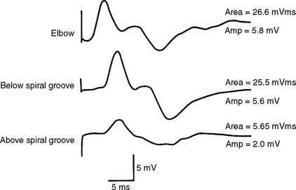

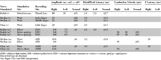
1. A partial right radial motor conduction block across the spiral groove, as evidenced by a drop in amplitude of the compound muscle action potential (CMAP) from 5.6 mV, stimulating below the spiral groove, to 2.0 mV, stimulating above the spiral groove (Figure C12-1). This 64% amplitude decay is supported by the lack of significant CMAP dispersion and the concomitant decrease in negative CMAP area from 25.5 mV/ms to 5.65 mV/ms, respectively (78% area loss). Also, there is relative and mild focal slowing of the conducting radial motor fibers within the spiral groove (when the right radial motor nerve conduction velocity of 57 m/s is compared to the distal velocity of 66 m/s, and to the left radial motor velocities of 69 m/s proximally and 68 m/s distally). The presence of partial conduction block with relative mild focal slowing across the spiral groove is consistent with segmental demyelination at that site.


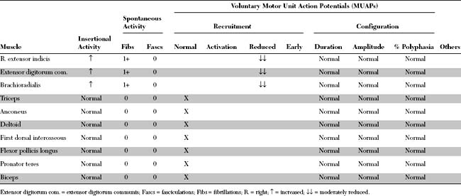
This case is consistent with a right radial mononeuropathy at the spiral groove, manifested mostly by segmental demyelination (partial conduction block within the spiral groove) with modest sensory and motor axonal loss. The presence of wristdrop and fingerdrop with weak brachioradialis but normal triceps and deltoid, along with superficial radial sensory deficit, makes the clinical diagnosis of radial nerve lesion in the region of the spiral groove very likely. Identifying a conduction block across the spiral groove localizes the lesion precisely to that segment of the radial nerve. In addition, the lesion could not be due to a posterior interosseous neuropathy because the radial sensory SNAP and brachioradialis muscle are abnormal; the motor branch to the brachioradialis (and the branch to the extensor carpi radialis longus) originates from the main trunk of the radial nerve before it divides into its terminal branches (posterior interosseous and radial cutaneous). In a posterior cord brachial plexus lesion, the deltoid, triceps, and anconeus muscles are abnormal. Finally, the SNAPs are normal in cervical radiculopathy (because the root lesion is proximal to the dorsal root ganglia), and muscles innervated by other nerves that share the same root should be affected.


In the case presented, the NCSs were done 5 weeks after the onset of the patient’s symptoms, long after the time required for wallerian degeneration (10–11 days). Thus, the conduction block seen cannot be due to early axonal loss, and the primary pathophysiologic process here is focal demyelinative block. Conduction slowing also is caused by demyelination and can accompany conduction block, although they often occur independently. Sensory and motor axonal degeneration has occurred, as confirmed by low-amplitude distal radial CMAP, low-amplitude distal radial SNAP, and fibrillation potentials in radial innervated muscles. The prognosis for this patient should be good but biphasic because it is dependent on the relatively rapid remyelination process and slower reinnervation. Reinnervation in this case should be efficient because the lesion is partial and sprouting is likely to be vigorous.


DISCUSSION


Applied Anatomy


The radial nerve is the largest nerve in the upper extremity (Figure C12-2). It is a direct extension of the posterior cord of the brachial plexus, after takeoff of the axillary nerve, and contains fibers from all the contributing roots of the plexus (i.e., C5 through T1).
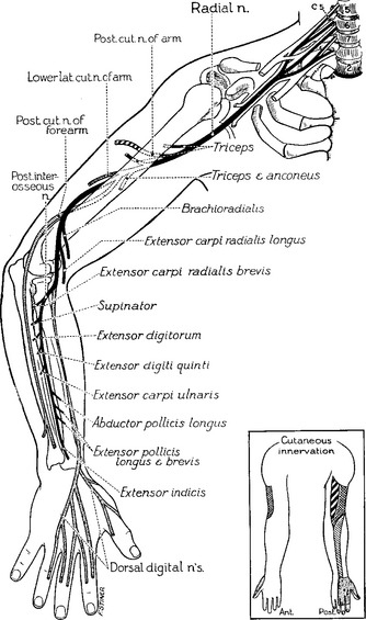


To attain better understanding of the anatomy and innervation of this long and serpiginous nerve, its path is best dissected into multiple segments:


Stay updated, free articles. Join our Telegram channel

Aug 12, 2016 | Posted by in CARDIOLOGY | Comments Off on Case 12

Full access? Get Clinical Tree

Get Clinical Tree app for offline access