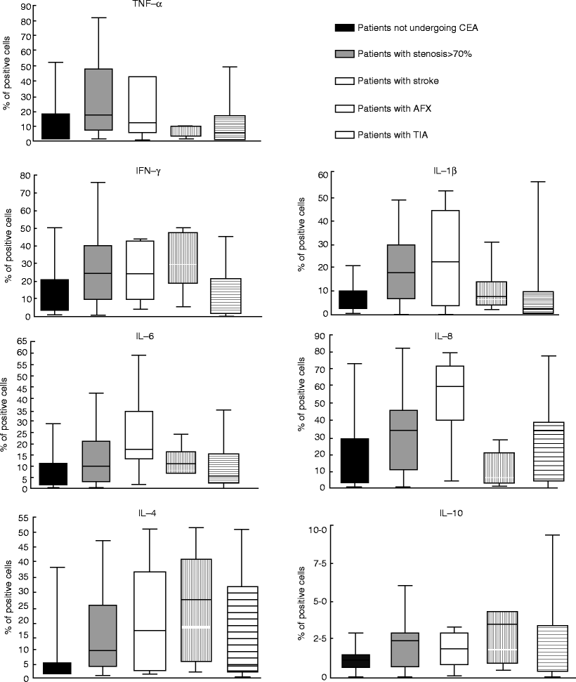
When studying the presence of inflammatory cytokines in patients undergoing CEA, intracellular cytokine expression was found to be significantly higher in patients undergoing CEA who had stenosis ≥70 % (TNF-α, IFN-γ, IL-1b, IL-6, IL-4, and IL-10), with previous stroke (IFN-γ, IL-1b, IL-6, IL-8, IL-4, and IL-10), and with amaurosis fugax (IFN-γ, IL-6, IL-4, and IL-10) than in patients not undergoing CEA (Table 5.2) [14]. Proinflammatory cytokines, such as TNF-α, IFN-γ, IL-1b, IL-6, and IL-8, and antiinflammatory cytokines, such as IL-4 and IL-10, were also found in significantly higher percentages in patients undergoing CEA than in patients who were not [3].

Table 5.2
Box-plot graphs showing medians and interquartile ranges of intracellular cytokines in the 39 patients not undergoing carotid endarterectomy (CEA) and in the 56 patients undergoing CEA divided according to the indications for surgery [18 asymptomatic patients with stenosis ≥70 %, 14 patients with stenosis <70 % and stroke, 7 patients with stenosis <70 % and amaurosis fugax (AFX), 17 patients with stenosis <70 % and transient ischemic attack (TIA)]. Patients with stenosis of 70 % versus patients with stenosis of <70 %: tumor necrosis factor (TNF)-a and interleukin (IL)-6, P = 0.004; interferon (IFN)-g and IL-10, P = 0.01; IL-1b, P = 0.02; IL-4, P = 0.006. Patients with stroke versus asymptomatic patients: IFN-g, P = 0.005; IL-1b and IL-4, P = 0.004; IL-6, P <0.0001; IL-8, P = 0.0001; IL-10, P = 0.03. Patients with AFX versus asymptomatic patients: IFN-g, P = 0.007; IL-6 and IL-10, P = 0.03; IL-4, P = 0.003

Significantly higher levels of IL-8-positive cells were observed in patients with contralateral than in those with monolateral carotid disease, demonstrating an association of IL-8-positive cells with the onset or progression of contralateral disease after CEA [13, 14].
The higher pro- and antiinflammatory cytokine expression in patients with stenosis of ≥70 % than in those with stenosis of <70 % could indicate plaque instability and immunological mechanisms leading to atherosclerotic plaque growth. However, the question of whether inflammatory cytokine expression has a direct causal relation with carotid arterial disease or only reflects the inflammatory disease process remains unanswered.
Gender Differences in Carotid Plaques
In their study on sex differences in the composition of plaques in patients undergoing CEA, Rerkasem et al. found that plaques from females had significantly less lipid and more fibrous tissue than those from males, and the perioperative stroke and death rate in females and males was 3.3 and 3.6 %, respectively [15]. This might reflect the fact that women have a more stable plaque component than men because unstable carotid plaques have a high lipid content, thin fibrous caps, a high inflammatory cell content, and increased protease activity.
Halvorsen et al. also supported the notion that women have more stable plaques with an ultrasound study on carotid arteries that showed that women had significantly fewer echolucent plaques than men [16].
Hellings et al. observed that the differences in plaque histology between men and women were paralleled by the inflammatory and protease activity in atherosclerotic plaques (Table 5.2). Women showed lower IL-8 values than men. IL-6 levels were not different between men and women. Protease activity was lower in women, with matrix metalloproteinase-8 showing significantly lower values than in men. Asymptomatic women showed lower levels of IL-8, matrix metalloproteinase-8 activity, and matrix metalloproteinase-9 activity compared with asymptomatic men [17].
Previous studies have shown that the presence of these matrix metalloproteinases plays an important role in cell migration, degradation of the fibrous cap, expansive remodeling, and intraplaque neovessel formation. This might suggest that matrix metalloproteinase contributes to plaque instability [18, 19].
Conclusion
Investigation of inflammatory cytokine provides an insight into the inflammatory mechanisms related to carotid atherosclerosis. However, a still unanswered question is whether cytokine expression in the peripheral blood has a causal relation with carotid atherosclerosis or simply reflects the inflammatory disease process. Studies should be performed on this issue, and in the future they will lead to improved therapeutic protocols and reduced atherosclerosis-related mortality.
References
1.
< div class='tao-gold-member'>
Only gold members can continue reading. Log In or Register to continue
Stay updated, free articles. Join our Telegram channel

Full access? Get Clinical Tree


