Carotid Artery Stenting

15 Carotid Artery Stenting


Nearly 800,000 strokes occur each year in the United States, and more than 130,000 Americans die annually from stroke. Stroke is the third leading cause of mortality in the United States, and among survivors, 15% to 30% are permanently disabled. Warning symptoms, such as a transient ischemic attack (TIA), historically defined as a neurologic event lasting <24 hours, precede a minority (15%) of all strokes. A new, tissue-based definition of TIA has been formalized, but most of the studies referred to in this chapter predate its publication. Following a TIA, the 90-day risk of stroke is 15%, and the 6-month risk of a stroke, TIA, or death is as high as 30%. Therefore, TIAs should be treated as medical emergencies.


Approximately half of all strokes occur in the distribution of the carotid artery, and carotid artery disease (CAD) amenable to revascularization accounts for as high as 12% of new strokes. Although occlusion of the carotid artery due to plaque burden can cause a stroke, the more common scenario is for carotid plaque to rupture resulting in distal embolization and cerebral infarction. Because more than 80% of strokes have no warning symptoms, stroke prevention with management of asymptomatic carotid atherosclerosis and carotid revascularization for high-risk patients is important.


Roughly 5% to 10% of patients over age 65 have a carotid stenosis >50%, and only 1% have a stenosis ≥75%. The natural history of carotid artery disease depends on the patient’s symptomatic status and lesion severity. In the Asymptomatic Carotid Atherosclerosis Study (ACAS), among medically treated asymptomatic patients with stenosis ≥60%, the 5-year risk of ipsilateral stroke or any stroke or death within 30 days of randomization was 11%.


Symptomatic patients with carotid atherosclerosis have a much worse prognosis than asymptomatic patients. In the North American Symptomatic Carotid Endarterectomy Trial (NASCET) trial of symptomatic carotid lesions, the 5-year risk of ipsilateral stroke in those medically managed was 18.7% among those with lesions <50% in severity. Results were similar in the European Carotid Surgery Trial (ECST). The risk of stroke increased with severity of stenosis, with the 3-year risk of ipsilateral stroke in those with stenosis >80% being 26.5%.


Medical therapy for carotid atherosclerosis should focus on preventing stroke and stabilizing atherosclerotic lesions to prevent plaque rupture and atheroembolization. Blood pressure control with angiotensin-converting enzyme inhibitors and angiotensin receptor blockers are of particular benefit in stroke prevention. In addition, a review of more than 70,000 patients with, or at high risk for, cardiovascular disease found that statins significantly lower the risk of stroke. Current American Heart Association/American Stroke Association (AHA/ASA) stroke guidelines endorse the National Cholesterol Educational Program (NCEP III) recommendations for the use of statins. The Justification for the Use of Statins in Prevention: An Intervention Trial Evaluating Rosuvastatin (JUPITER) study showed that rosuvastatin treatment in patients with normal cholesterol levels but elevated levels of C-reactive protein is effective in reducing the rate of stroke and is indicated for patients with carotid artery disease.


Antiplatelet medications are a critical component of primary stroke prevention. In secondary prevention, aspirin reduces the risk of future strokes by 15% to 25%. High-dose aspirin provides no more benefit than lower doses (160–325 mg daily) but is associated with more side effects. The Clopidogrel for High Atherothrombotic Risk and Ischemic Stabilization, Management, and Avoidance (CHARISMA) trial included more than 4300 patients with a prior TIA or stroke and found that aspirin 75 to 162 mg daily was as effective as aspirin plus clopidogrel in preventing future myocardial infarction (MI), stroke, or cardiovascular death. AHA/ASA guidelines recommend that all patients with carotid atherosclerosis be placed on antiplatelet medications. Aspirin 50 to 325 mg daily, aspirin-dipyridamole, or clopidogrel should be initiated for secondary prevention of stroke. The combination of aspirin and dipyridamole is recommended over aspirin alone, however. The combination of aspirin and clopidogrel is not recommended.


Diagnosis of Carotid Artery Disease: Anatomical Imaging


Duplex ultrasonography is most often the initial test used to assess the severity of carotid artery stenosis. Carotid ultrasound also has a high accuracy for carotid restenosis after endarterectomy. Numerous criteria have been proposed to diagnose severe carotid stenosis. In most cases >80% stenosis correlates with systolic velocity >300–400 cm/sec, diastolic velocity >100–135 cm/sec, and ratio of internal carotid artery/common carotid artery (ICA/CCA) systolic velocity of >3.5. Factors such as contralateral occlusion, diminished cardiac output from severe left ventricular dysfunction, aortic stenosis, and common carotid artery stenosis may make these measurements less reliable. Magnetic resonance angiography and computed tomography angiography are the other noninvasive imaging studies that are helpful in identifying carotid artery stenosis. Duplex imaging is often the test of choice given its safety profile, low cost, and wide availability. Imaging that can define the aortic arch and the circle of Willis is useful in planning endovascular procedures. The particular noninvasive method used should reflect local availability and expertise.


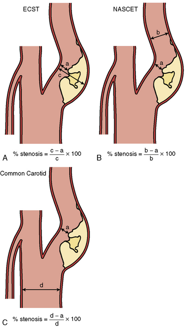
Digital subtraction angiography (DSA) is the gold standard for defining carotid anatomy with the NASCET method of stenosis measurement the most widely used (Fig. 15-1). Cerebral catheter-based angiography carries a risk of cerebral infarction of 0.5% to 1.2%; therefore, noninvasive imaging should be the initial strategy for evaluation.


Procedural Techniques


Baseline Aortography and Cerebral Angiography


Vascular access is most commonly obtained from the femoral artery although brachial or radial access may be used. Prior to selective angiography, an arch aortogram is performed with a pigtail catheter placed in the proximal ascending aorta to define the anatomy of the aortic arch, which is critical to the success of the stent procedure. This is done in the 45º left anterior oblique position with a large-format image intensifier (12-inch to 16-inch) using DSA (15 mL/sec for 3 sec) and a power injector.


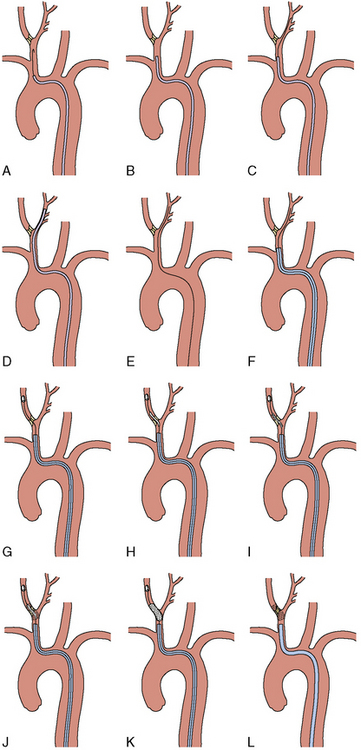
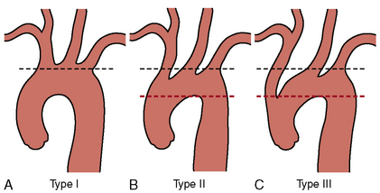
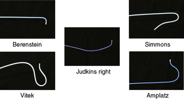
Once the morphology of the aortic arch is determined (Fig. 15-2), catheters are chosen for selective angiography of the cervical arteries supplying the brain (right and left carotid and vertebral arteries) and the cerebral vasculature. In a type I arch, Berenstein or Judkins right (JR) catheters are often used. In type II or III arch morphologies, shepherd’s crook–shaped catheters (i.e., Simmons or Vitek catheters) may be best (Fig. 15-3).


Angiograms are obtained to delineate the anterior and posterior circulation supplying the brain. The intracranial and extracranial portions of each vessel are studied. Generally, two views of each are obtained, one in the anteroposterior projection and one in the lateral projection. Alternatively, some operators use rotational angiography. It is important to demonstrate the circle of Willis to define any baseline abnormalities. DSA may be performed with a 50/50 mix of saline and contrast. An external reference object is used with carotid angiograms in order to accurately measure the diameter of the artery.


Internal Carotid Intervention


A diagnostic catheter is used to engage the common carotid artery (CCA), and a roadmap angiogram is made of the carotid bifurcation. A 0.035-inch stiff-angled hydrophilic wire is advanced into the external carotid artery, and the diagnostic catheter is advanced over the wire. The hydrophilic wire is exchanged for a 0.035-inch stiff Amplatz wire over which an 8F guiding catheter or a 6F sheath may be advanced to the target vessel CCA. Care must be taken to avoid plaque disruption with wires and catheters, and at this point in the procedure, the plaque in the ICA should remain untouched.


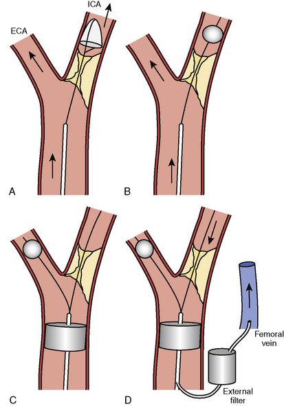
For procedures performed with a filter-type distal embolic protection device (EPD), the target lesion is crossed with the EPD. Although there are no randomized trials comparing stenting with EPDs to stenting alone, one study found that 57% of EPDs contained debris upon retrieval. EPDs are standard of care in the United States, and several types exist (Fig. 15-4). If the distal EPD will not cross the lesion, the stenosis may be crossed with a conventional 0.014-inch guidewire and subsequently predilated with a small (2.5 mm) balloon. Then the EPD should be placed. After distal EPD deployment, the lesion is often predilated with an undersized coronary balloon, typically 3 to 4 mm in diameter. A self-expanding stent is then placed across the lesion. The stent is sized to fit the CCA, and as a general rule, self-expanding stents are typically sized at least 1 mm larger than the reference diameter. There is no demonstrated benefit for using tapered stents. It is common practice, when treating an internal carotid bifurcation lesion, to place the stent across the ostium of the external carotid artery (Fig. 15-5).


Jun 5, 2016 | Posted by in CARDIAC SURGERY | Comments Off on Carotid Artery Stenting

Full access? Get Clinical Tree

Get Clinical Tree app for offline access