Carotid Artery Disease Management


Carotid Artery Disease Management


Debabrata Mukherjee

Aamer Abbas


In western countries, stroke affects approximately 0.2% of the population annually. In the United States alone, around 500,000 people experience a new stroke and 200,000 have a recurrence of a previous cerebrovascular event each year. About 90% of the strokes are ischemic and 10% are hemorrhagic (intracerebral hemorrhage or subarachnoid hemorrhage). The major cause of stroke is large-vessel atherosclerosis, with the highest risk occurring in patients with stenosis of the internal carotid arteries. Among patients who have suffered a stroke, one third may die within 1 year, one third may have permanent disability, and the remainder recovers. Stroke is the third leading cause of death in Western countries, accounting for 12% of all deaths. According to the data from Health Care Financing, in 1996, $3.8 billion ($5,945 per discharge) was paid in the United States to Medicare beneficiaries for stroke. The total stroke-related burden for the American economy is estimated to be $20 billion yearly due to health care costs and lost productivity. Stroke thus remains a major public health problem.

The classical risk factors of coronary atherosclerosis also apply to carotid atherosclerosis. Accordingly, presence and severity of carotid atherosclerosis correlate with the presence and severity of coronary atherosclerosis and peripheral vascular disease. Half of the men over 75 years of age have carotid atherosclerosis by ultrasonography, with a stenosis greater than 50% detected in 5% of cases. Important risk factors for carotid stenosis include smoking, diabetes, male gender, hypertension, and dyslipidemia. In patients with carotid atherosclerosis on ultrasound but no neurologic symptoms, the risk of subsequent stroke is best predicted by the percent carotid stenosis, the presence of disease progression on sequential ultrasound examinations, and the presence of carotid ulceration. Recent data demonstrated a risk of 1.6% per year on aspirin among asymptomatic patients with carotid stenosis less than 60% and of 3.2% per year among those with stenosis greater than 60%. Among patients with symptomatic [i.e., with previous stroke or transient ischemic attack (TIA)] carotid disease, the risk of recurrence may be as high as 10% in the first year and 30 to 35% at 5 years. High-risk features for recurrence include hemispheric TIA, recent TIA, increasing frequency of TIA, or high-grade carotid stenosis.

The most common site of cerebrovascular atherosclerotic disease is the carotid bifurcation, characteristically affecting the outer wall of the carotid sinus and extending into the distal common carotid artery. Atherosclerotic plaque rupture with subsequent total thrombotic occlusion of the carotid is a rare phenomenon. More commonly, atherosclerotic
ulceration leads to luminal thrombus apposition and distal embolization. Both in asymptomatic and in symptomatic patients, the more severe the carotid lesion, the higher the risk of subsequent stroke. The presence of ulcerated lesions, seen in up to one third of the endarterectomy specimen but underappreciated with angiography, also increases the risk of ischemic events. In the medical arm of the North American Symptomatic Carotid Endarterectomy Trial (NASCET), the 2-year stroke incidence among symptomatic patients with evidence of carotid ulcers on angiography ranged from 26.3 to 73.2% as the degree of stenosis increased from 75 to 95%.


CLINICAL FEATURES

The majority of patients with carotid stenosis are asymptomatic, and diagnosis is made following auscultation of a carotid bruit or routine ultrasound screening. TIA is the leading symptom, and in the absence of neurologic deficits, the diagnosis is based on careful history taking. If no therapy is instituted, as many as 30 to 40% of these patients may subsequently develop stroke. Dysphasia, ipsilateral amaurosis fugax, contralateral visual field, and motor and sensory loss may be all manifestations of carotid disease. These findings must be differentiated from vertebrobasilar disease. While neurologic findings associated with carotid disease usually involve the contralateral face and body, posterior circulation events often cause bilateral or crossed deficits such as ataxia, dysarthria, diplopia, or bilateral visual field loss. Headache is an uncommon finding associated with cerebral embolic events. Hemicranial headache may occur with TIA originating from carotid stenosis, and occipital headaches may be reported in vertebrobasilar insufficiency. In patients with neurologic deficits associated with neck or retro-orbital pain, carotid or vertebral artery dissection should be excluded, particularly in the presence of trauma. The clinical triad of neck, head, or retro-orbital pain; new-onset Horner’s syndrome; and contralateral sensory, motor, or cognitive deficits is pathognomonic for internal carotid artery (ICA) dissection.


DIAGNOSIS


Ultrasonography

Duplex ultrasonography, a combination of Doppler ultrasonography and B-mode imaging generated by a single transducer, is the diagnostic tool of choice for the initial assessment of carotid disease. This widely available technique allows morphological and functional assessment of the carotid lesion. When performed by trained sonographers using a standard protocol and with ongoing quality assurance, this method approaches 90% sensitivity and specificity compared with angiography for detection of severe carotid stenosis. Percent stenosis is determined by systolic and diastolic velocities with peak end-diastolic velocity greater than 135 cm/s and peak end-systolic velocity greater than 240 cm/s suggestive of stenosis greater than 80%. Duplex carotid scans may yield inadequate images in the following
conditions: carotids that bifurcate high, long (greater than 3 cm) ICA plaque, calcific shadows, or near-complete occlusions. In these cases, magnetic resonance imaging is of value in differentiating between the two.


Magnetic Resonance Angiography

Magnetic resonance angiography (MRA) is a useful tool in carotid disease, allowing reliable imaging from the aortic arch to the intracranial branches. MR techniques for the diagnosis of carotid disease include gadoliniumbased (contrast enhanced—CE) and time-of-flight (TOF-flow sensitive) approaches. CE-MRA techniques have been shown to have high sensitivity (greater than 90%), high negative predictive value (greater than 90%), and high specificity (greater than 90%) for detecting severe carotid stenosis. On the other hand, TOF (2D and 3D) techniques may be compromised by artifacts generated by vessel tortuosity, blood flow turbulence, and in-plane flow, and it is precisely in these situations that CE-MRA methodology may be superior. TOF is also time-consuming and subject to movement artifact. The specificity of CE-MRA is at least equal to and may even exceed that of duplex ultrasound in the diagnosis of 70 to 99% lesions and in the diagnosis of subtotaled arteries versus complete occlusions. In situations where duplex ultrasound yields suboptimal results, MRA may help resolve the issue and reduce the need for an invasive assessment [high bifurcation, long (greater than 3 cm) ICA plaque, calcific shadows]. MRA also provides the advantage that concomitant brain imaging may be performed to assess the extent of ischemic events and to exclude intracranial pathologies (e.g., tumors or vascular malformations) prior to endovascular therapy.


CT Angiography

MDCT angiography is an important diagnostic modality to assess the carotid circulation. The intracranial as well as extracranial circulation can be evaluated in the same sitting with this modality. In a study of 37 patients and 73 vessels, the reported sensitivity and specificity for highgrade stenosis were 75 and 96, respectively, and for moderate stenosis 88 and 82, respectively, as compared to contrast angiography. Furthermore, MDCT may assess the composition of the atherosclerotic plaque and the hemodynamics of the brain circulation by using the CT brain perfusion technology.


Angiography

Angiography remains the gold standard for diagnosis of carotid stenosis. It allows accurate measurement of luminal stenosis of the entire vessel from its origin to the intracranial branches, as well as proper assessment of plaque morphology, lesion length, and reference vessel diameter. In patients with symptomatic carotid disease, angiography may identify additional intracranial stenosis. However, it is an invasive procedure with a potential for embolization or vessel trauma. Currently, angiography is used in patients where noninvasive tests yield inconclusive or conflicting results and/or when percutaneous therapy is considered. Two different
methods for calculating the degree of angiographic stenosis have been used in the major clinical trials (Fig. 11.1). Both the NASCET and the European Carotid Surgery Trial (ECST) define the stenotic segment the same way but differ in how they define the reference segment or the normal diameter. The NASCET method defines normal as the diameter just distal to the carotid bulb (neither the bulb itself nor a region of poststenotic dilatation), whereas the ECST method defines normal as the estimated diameter of the carotid bulb as it was prior to atherosclerotic narrowing. For any given stenosis, the ECST method gives a higher percentage diameter stenosis as illustrated in Figure 11.1. It is important to know which method was used to determine reference segment in order to interpret and apply trial results appropriately. Importantly, angiography remains the only scientifically validated method of defining ICA stenosis requiring endarterectomy, as in the large-scale randomized trials (i.e., NASCET, ECST) the degree of stenosis was assessed by angiography. However, in centers where MRA and duplex ultrasound imaging have been demonstrated to be concordant with angiography, angiography is usually not considered to be necessary in the clinical assessment of carotid disease prior to endarterectomy.


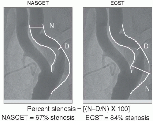
FIGURE 11-1. Carotid stenosis severity measurements according to the NASCET and the ECST methods. For any given stenosis, the ECST method gives a higher percentage diameter stenosis. (N, presumed normal segment; D, diseased segment) (Adapted from Mukherjee D, Yadav SJ. Cerebrovascular diseases: pathophysiology and management. In: Fuster V, ed. Assessing and modifying the vulnerable atherosclerotic plaque. Armonk, NY: Futura Publishing, 2002:29-55.)


MANAGEMENT OF CAROTID DISEASE


Risk Factor Modification and Antiplatelet Therapy


Hypertension Control

Hypertension has a strong correlation with stroke, with both systolic and diastolic blood pressures being independently associated with primary and recurrent stroke risk. Diastolic pressures are correlated strongly with risk in younger individuals, and a reduction in diastolic blood pressure of 1 mm Hg has been shown to decrease the incidence of stroke by 7 to 10% over 5 years. In older individuals, a reduction in systolic pressure of 2 mm translates into a reduction of stroke incidence of 5 to 10%.


Smoking Cessation

Cigarette smoking increases the risk of stroke two- to threefold, especially in conjunction with hypertension. Smoking cessation is an essential part of stroke management.


Diabetes

Diabetes increases the risk of stroke 3- to 10-fold depending on the age of presentation. Diabetes doubles the rate of recurrent stroke, trebles the frequency of stroke-related dementia, and also increases stroke-related mortality. However, there are as yet no good data showing that tight glycemic control reduces stroke rates in diabetic individuals. Preliminary studies suggest that insulin sensitizers may have beneficial effects in reducing carotid atherosclerosis and intimal-medial thickening and may translate into reduction in strokes.


Lipid Lowering

The Stroke Prevention by Aggressive Reduction in Cholesterol Levels (SPARCL) trial was a randomized, double-blind study designed to determine whether atorvastatin 80 mg/d or placebo would reduce the risk of fatal or nonfatal stroke in patients with no known coronary disease who had experienced a stroke or TIA within the previous 6 months. During a median follow-up of approximately 5 years, 11.2% receiving atorvastatin and 13.1% receiving placebo reached the primary end point of fatal or nonfatal stroke (5-year absolute reduction in risk, 2.2%; adjusted HR, 0.84; 95% confidence interval, 0.71, 0.99; P = 0.03). The AHA/ASA recommendations state that on the basis of the SPARCL trial, administration of statin therapy with intensive lipid-lowering effects is recommended for patients with atherosclerotic ischemic stroke or TIA and without known CHD to reduce the risk of stroke and cardiovascular events.


Antiplatelet Therapy

Table 11.1 summarizes the data on antiplatelet therapy in high-risk individuals with a focus on stroke as a primary or a secondary end point. In the ASA in Carotid Endarterectomy (ACE) trial, low-dose ASA (75 to


325 mg) was superior to high-dose ASA (650 to 1,300 mg) in preventing stroke, myocardial infarction (MI), or death both at 30 days (5.4 vs. 7.0%, P = 0.07) and at 3 months (6.2 vs. 8.4%, P = 0.03) in patients undergoing carotid endarterectomy. The Antiplatelet Trialists’ Collaboration analyzed the results of 195 trials of greater than 135,000 patients and found that platelet antagonists lowered the risk of stroke, MI, and vascular death. On the basis of this, individuals at risk for vascular events should receive antiplatelet agents for the primary prophylaxis of stroke.


TABLE 11.1 AGENTS EFFECTIVE IN VASCULAR RISK REDUCTION INCLUDING ONGOING CLINICAL TRIALS


Agent


Trials


Trial Specifics


N


1° End point (Reduction in Vascular Death/MI/Death)


Reduction in Nonfatal Stroke


Aspirin


I° Prevention


Physicians Health Study British Physicians Health Study


DBPC trials, ASA 325 mg vs. placebo


22,071 5,139


41% RR ↓ in MI (limited to >50 y)


NS ↑ in hemorrhagic stroke


II° Prevention


Meta-analysis


Antithrombotic Trialist’s Collaboration


>110,000


19%


25% RR ↓ with ASA compared to placebo in nonfatal stroke


II° Prevention, patients scheduled for endarterectomy


ACE


DBPC, Aspirin 81 mg vs. 325 mg vs. 650 mg or 1,300 mg for 3 mo


2,849


Rate of stroke, MI, and death was lower in the low-dose groups than the high-dose groups at 30 d (5.4 vs. 7.0%) and at 3 mo (6.2 vs. 8.4%)


Strokes occurred in 3-2% patients on low-dose ASA and 6-9% patients on highdose acetylsalicylic acid


Clopidogrel


II° Prevention High-risk patients with prior MI, stroke, or PAD


CAPRIE


DBPC, Clopidogrel 75 mg vs. ASA 325 mg


19,185


Statistically significant 8.7% RR ↓ in stroke, MI, or vascular death


Trend in reduction of stroke in subgroup with prior stroke


II° Prevention Patients with acute coronary syndrome


CURE


DBPC, Aspirin 75-325 mg vs. ASA + c lopidogrel 75 mg


12,562


20% RR ↓ in (11.4% in placebo vs. 9.3% in clopidogrel arm) stroke, MI, or vascular death


14% RR ↓ in stroke with clopidogrel


II° Prevention Patients with acute coronary syndrome


CREDO


DBPC, Clopidogrel 75 mg for 4 wk vs. clopidogrel 75 mg for up to 9 mo


2,116


26.9% RR ↓ in (11.5 vs. 8.5% in long-term clopidogrel arm) of death, MI, and stroke


No ↑ in hemorrhagic strokes


II° Prevention Patients with recent TIA or stroke


MATCH


DBPC, Clopidogrel 75 mg vs. ASA + clopidogrel 75 mg


7,600


15.7% reached the primary end point in the group receiving aspirin + clopidogrel vs. 16.7% in the clopidogrel alone group [relative risk reduction 6.4%, (95% CI 4.6-16.3)]; Life-threatening bleedings were higher in the group receiving aspirin + clopidogrel vs. clopidogrel alone [96 (2.6%) vs. 49 (1.3%); absolute risk increase 1.3% (95% CI 0.6-1.9)]


No benefit with dual therapy but ↑ bleed


II° Prevention Patients with small-vessel disease and lacunar infarcts


SPS3


DBPC, Aspirin 325 mg vs. aspirin 325 mg + clopidogrel 75 mg


2,500


Phase III trial ongoing


Trial ongoing


II° Prevention Patients who have recently recovered from a TIA


PRoFESS


DBPC, aspirin + extended-release dipyridamole vs. clopidogrel monotherapy


20,332


Recurrent stroke 1.01 (0.92-1.11); the secondary outcome, a composite of stroke, MI, and vascular death, was identical between groups


Neither aspirin plus extended-release dipyridamole or clopidogrel is superior to the other in the prevention of stroke


Dipyridamole


II° Prevention Patients with TIA or stroke within 3 mo


ESPS-2


DBPC, 2×2 factorial design, ASA 25 b.i.d., dipyridamole (ER-DP) 200 b.i.d.; placebo and ASA + ER-DP


6,602


No effect on MI or mortality; stroke rate at 24 mo — 9.5% with aspirin + dipyridamole vs. 12.5% with aspirin alone


37% reduction in stroke with aspirin + dipyridamole vs. 19% reduction with aspirin alone Dose of aspirin used was only 50 mg


II° Prevention Patients with TIA or stroke within 3 mo


ESPRIT


DBPC, aspirin (30-325 mg daily) vs. aspirin + dipyridamole (200 mg twice daily)


2,739


Hazard ratio with combination 0.80, (95% CI 0.66-0.98); absolute risk reduction 1.0% per year, (95% CI 0.1-1.8)


Combination regimen of aspirin plus dipyridamole is preferred over aspirin alone as antithrombotic therapy after cerebral ischemia of arterial origin


HMG CoA Reductase Inhibitors (Statins)


Patients with average cholesterol levels with prior MI


CARE


DBPC, randomized to pravastatin or placebo


4,159


24% reduction in heart attack or death


31% reduction in stroke


Patients with a history of heart attack or hospitalization for unstable angina pectoris


LIPID


DBPC, randomized to pravastatin or placebo


9,014


24% reduction in heart attacks, and a 22% decrease in deaths from any cause


19% reduction in stroke


Patients with acute coronary syndrome


MIRACL


DBPC, randomized to atorvastatin or placebo


16% RR ↓ in 14.8% in the atorvastatin group and 17.4% in the placebo group


50% reduction in stroke


People who have a high risk of coronary artery disease—even if their cholesterol levels are normal or low


HPS


DBPC, randomized to simvastatin or placebo


20,000


25% reduction in stroke


2 secondary prevention studies, 5 mixed primary-secondary prevention population studies, and 10 regression trials


Meta-analysis of 17 trials


Meta-analysis of all randomized controlled trials published as of April 1997


21,303


24% RR ↓ in all-cause mortality


31 % reduction in stroke


Patients who had had a stroke or TIA within 1-6 mo before study entry


SPARCL


DBPC, randomized to atorvastatin or placebo


4,731


During a median follow-up of 4.9 y, 11.2% receiving atorvastatin and 13.1% receiving placebo had a fatal or nonfatal stroke [5-y absolute reduction in risk, 2.2%; adjusted hazard ratio, 0.84; (95% CI 0.71-0.99; P= 0.03); unadjusted P= 0.05]


Atorvastatin reduced the overall incidence of strokes and of cardiovascular events by 16%


DBPC, double-blind placebo controlled trial; RR, relative risk; PAD, peripheral arterial disease; TIA, transient ischemic attack; ACE, ASA and carotid endarterectomy; CAPRIE, clopidogrel vs. aspirin in patients at risk of ischemic events; CURE, clopidogrel in unstable angina to prevent recurrent events; CREDO, clopidogrel for the reduction of events during observation; MATCH, management of atherothrombosis with clopidogrel in high-risk patients with recent TIA or ischemic stroke; SPS3, secondary prevention of small subcortical stroke; PRoFESS, prevention regimen for effectively avoiding second strokes; ESPRIT, European/Australian stroke prevention in reversible ischemia trial; ESPS, European stroke prevention study; CARE, cholesterol and recurrent events; LIPID, long-term intervention with pravastatin in ischemic disease; MIRACL, myocardial ischemia reduction with aggressive cholesterol lowering; HPS, Heart protection study; SPARCL, stroke prevention by aggressive reduction in cholesterol levels.


The efficacy of dual antiplatelet therapy with aspirin and clopidogrel was tested in the Management of Atherothrombosis with Clopidogrel in High-Risk Patients with Recent TIA or Ischemic Stroke (MATCH) trail. The MATCH trial did not show additional clinical value of adding aspirin to clopidogrel in high-risk patients with TIA or ischemic stroke. The European/Australian Stroke Prevention in Reversible Ischemia Trial (ESPRIT) was a randomized, open-label study comparing aspirin 30 to 325 mg with or without dipyridamole 200 mg b.i.d. in 2,763 subjects with TIA, transient monocular blindness, or minor stroke within 6 months of enrollment. The risk for the primary outcome of death from all vascular causes, nonfatal stroke, nonfatal MI, or major bleeding complication was significantly lower in the dipyridamole plus aspirin arm (HR, 0.80; 95% confidence interval, 0.66 to 0.98). The Prevention Regimen for Effectively Avoiding Second Strokes (PRoFESS) trial demonstrated that the risks of recurrent stroke or the composite of stroke, MI, or vascular death are similar with aspirin + extended-release dipyridamole combination and clopidogrel monotherapy. The update to the AHA/ASA recommendations for the prevention of stroke in patients with stroke and TIA recommends that aspirin (50 to 325 mg/d) monotherapy, the combination of aspirin and extended-release dipyridamole, and clopidogrel monotherapy are all acceptable options for initial therapy (Class I, Level of Evidence A). Based on ESPRIT trial, the combination of aspirin and extended-release dipyridamole is recommended over aspirin alone (Class I, Level of Evidence B).


Surgical Revascularization


Symptomatic Carotid Disease

The main findings of randomized trials comparing carotid endarterectomy with medical management are summarized in Table 11.2. Three pivotal studies in patients with symptomatic carotid disease have been completed and have documented improved outcomes with endarterectomy in patients with symptomatic severe carotid stenosis. The ECST trial was a multicenter, randomized trial in which patients with nondisabling stroke, TIA, or retinal infarction within the preceding 6 months were randomly assigned to carotid endarterectomy or medical therapy. The rate of perioperative major stroke or death was 7.0%. Patients with severe stenosis (greater than 70%) allocated to surgery had significant reduction in death or any stroke at 3-year follow-up (12.3 vs. 21.9%; P < 0.01). The risk of surgical death or ipsilateral stroke by 3 years was 10.3% in patients assigned to surgical intervention compared with 16.8% in patients assigned to medical therapy. Patients with mild and moderate carotid stenosis did not benefit with
surgery. The NASCET trial randomized patients with symptomatic 30 to 99% carotid artery stenosis to endarterectomy or medical management. Among 662 patients with 70 to 99% stenosis, at 2 years, those randomized to surgery had benefit in terms of ipsilateral stroke (9 vs. 26%; absolute risk reduction 17%; P < 0.001), any stroke (12.6 vs. 27.6%; P < 0.001), and major stroke or death (8.0 vs. 18.1%; P < 0.01). The perioperative (i.e., within 30 days) stroke or death rate in the trial was 5.8%. A total of 2,267 patients had stenosis less than 70% and were divided in post hoc analyses into groups with 50 to 69% stenosis and those with less than 50% stenosis. In patients with 50 to 69% stenosis, a 6.5% reduction in the

Only gold members can continue reading. Log In or Register to continue

Stay updated, free articles. Join our Telegram channel

Jun 12, 2016 | Posted by in CARDIOLOGY | Comments Off on Carotid Artery Disease Management

Full access? Get Clinical Tree

Get Clinical Tree app for offline access