Cardiovascular Disease in Pregnancy

58 Cardiovascular Disease in Pregnancy


As more women delay childbearing into their thirties and forties, the interaction between coronary disease, its risk factors, and pregnancy becomes increasingly important in prenatal care. In addition to traditional cardiovascular risk, more women with congenital heart disease are reaching childbearing age. Pregnancy presents unique challenges for the management of cardiovascular diseases, necessitating a multidisciplinary approach to achieve optimal maternal and fetal outcomes. Understanding normal physiologic adaptations to pregnancy and their potential effect on cardiovascular hemodynamics is central to the management of pregnant women with coronary artery, valvular, congenital, or myocardial abnormalities.


Physiologic Adaptations to Pregnancy


Changes during Pregnancy


Important hemodynamic changes occur during pregnancy as a result of increases in red blood cell mass and plasma volume. Red blood cell mass typically increases by 20% to 30%, while plasma blood volume can increase even more—generally by about 50%. The etiology of the increase in blood volume is multifactorial and due mainly to activation of the renin-angiotensin-aldosterone system by estrogen. In addition, other pathways responsible for water retention are stimulated by other pregnancy-related hormones (Fig. 58-1). This relative increase in total blood volume results in a relative anemia, referred to as the physiologic anemia of pregnancy.


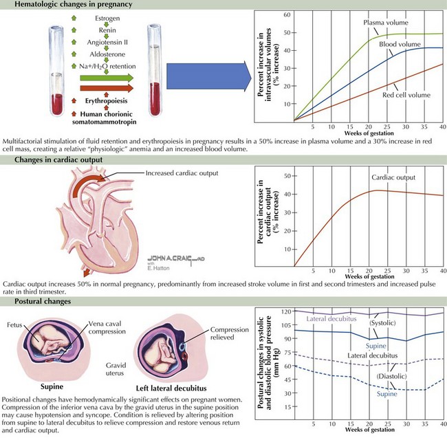
Cardiac output increases by approximately 45% during a normal pregnancy, starting as early as 5 weeks after the last menstrual period, predominantly from an increase in stroke volume (during the first and second trimesters) and an increase in heart rate (10–20 bpm during the third trimester). Most of the increase in cardiac output occurs by gestational week 16. This increase is followed by a further, slower increase in cardiac output that peaks at week 24 until week 32. Systemic vascular resistance (SVR) decreases 34% by 20 weeks as a result of decreased aortic compliance and arteriovenous shunting in the uterus. Subsequently, in the final weeks of pregnancy there is a slight decrease in cardiac output that reflects the decrease in stroke volume due to increased SVR (see Fig. 58-1, middle).


Related to these hemodynamic changes are structural changes of the heart. The left ventricular (LV) mass increases because of increased LV end-diastolic volume, decreased LV end-systolic volume, and increased wall thickness. The valvular cross-sectional area also increases, resulting in more physiologic regurgitation, affecting the tricuspid and pulmonary valves more commonly than the mitral valve. Although flow murmurs (due to increased flow across the aortic valve) are common in pregnancy, it is rare that there is sufficient tricuspid or pulmonic regurgitation to result in a murmur or significant hemodynamic effects.


Positional changes also have hemodynamically significant effects on the pregnant woman. Of particular importance is the supine hypotension syndrome characterized by symptoms of near-syncope/syncope caused by compression or occlusion of the inferior vena cava by the gravid uterus when the pregnant woman lies supine. Symptoms can be relieved by assuming another position, particularly the left lateral decubitus position (see Fig. 58-1, lower). The supine hypotension syndrome is one of the primary reasons to advise pregnant women against exercising in the supine position after the first trimester. This positional effect must also be recognized in the event that a pregnant woman (particularly in the second or third trimester) requires cardiopulmonary resuscitation. If this unfortunate situation arises, the woman should be placed in the left lateral decubitus position.


Clinical Presentation


Cardiac Examination during Normal Pregnancy


The symptoms of normal pregnancy—including fatigue, dyspnea, palpitations, and even near-syncope—in association with the normal signs of pregnancy (including augmentation of the jugular venous pulsations, normal heart sounds or murmurs, and a modest amount of lower extremity edema) may be misinterpreted as those of cardiac disease. Conversely, pathologic signs and symptoms at times may be attributed to normal pregnancy. Thus, knowledge of the normal cardiac examination during pregnancy is crucial (Table 58-1).


Table 58-1 Normal Physical Findings for the Cardiac Examination during Pregnancy


Examination Findings
Precordial palpation

Heart sounds


Heart murmurs


LV, left ventricular; RV, right ventricular.


Although the presence of an S3 sound is generally considered a normal finding in pregnancy and in young adults, it is still relatively rare in the healthy pregnant state. An S4 is unusual and generally indicates underlying cardiovascular pathology. Because of increased plasma volume and cardiac output, as noted previously, new or more prominent systolic flow murmurs are often present during pregnancy. Although diastolic murmurs have been reported in normal pregnancy, if a diastolic murmur is identified, further workup is indicated. Transthoracic echocardiography should be performed to evaluate for valvular pathology. With more plasma volume the pregnant woman may show mild jugular venous distention and peripheral edema. The pulse pressure will typically increase with more decrease in the diastolic blood pressure than in the systolic component.


Preexisting Disease States and Pregnancy


Maternal and fetal risks of cardiac disease generally depend on the underlying cardiovascular lesion and the functional class of the mother. Overall, women with functional New York Heart Association (NYHA) class I and II have a low mortality rate (<1% during pregnancy), whereas those with NYHA class III and IV have a much higher associated mortality rate (>7%). An updated risk index has been proposed to better risk-stratify pregnant women with heart disease. This index includes four predictors of primary events: prior cardiac events or arrhythmia; baseline NYHA class greater than II or cyanosis; significant left heart obstruction (mitral valve area <2 cm2, aortic valve area <1.5 cm2, or a peak LV outflow tract gradient >30 mm Hg by echocardiography); and reduced systemic ventricular systolic function (ejection fraction <40%).


Ideally, prepregnancy counseling provides the patient with information about case-specific maternal and fetal risks to prepare for the safest pregnancy possible. This also allows the physician and the patient to discuss risk factor modification and potential prenatal surgical correction of the underlying defect if pregnancy is desired.


Congenital Heart Disease


Congenital heart disease is thought to be multifactorial in origin, arising from a genetic predisposition combined with environmental factors. In general, the risk to offspring is modest (~3% to 5%) but much greater than in the general population. It should be noted, however, that reported rates vary between 1% and 18%, depending on the specific type of maternal lesion and the number of affected siblings. Maternal congenital heart disease confers different risks to both the mother and fetus, depending on the type of lesion (Table 58-2).


Table 58-2 Congenital Heart Disease and Maternal and/or Fetal Risk during Pregnancy (Excluding Valvular Disease)


High Risk Moderate Risk Low Risk


LV, left ventricular; NYHA, New York Heart Association; TGA, transposition of the great arteries.


Uncomplicated acyanotic lesions, including atrial and ventricular septal defects, patent ductus arteriosus (with left to right shunting), and aortic coarctation, are usually well tolerated during pregnancy. Patients with coarctation who develop severe hypertension are at risk for heart failure, cerebral aneurysm rupture, and aortic dissection. Therefore, modest, but not aggressive, blood pressure control is warranted for this population.


The maternal and fetal outcomes of pregnancy in acyanotic and cyanotic women with congenital heart disease are favorable provided that their NYHA functional class is I or II and the ejection fraction measured at the beginning of pregnancy is normal. However, the outcome of pregnant women with cyanotic or complex lesions depends significantly on the type of lesion, the state of surgical repair (if any), the degree of pulmonary hypertension, the magnitude of hypoxemia, and the functional status of the mother. Hence, it is important to address each case individually.


Jun 12, 2016 | Posted by in CARDIOLOGY | Comments Off on Cardiovascular Disease in Pregnancy

Full access? Get Clinical Tree

Get Clinical Tree app for offline access