Fig. 23.1
Chest x-ray at presentation showing enlarged heart and pulmonary oedema
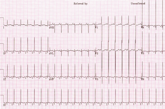
Fig. 23.2
ECG at presentation with inferior and lateral P wave inversion. The heart rate was 163/min
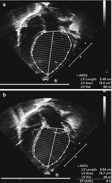
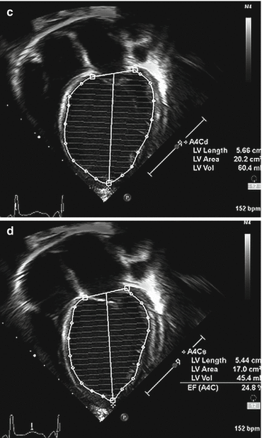
Fig. 23.3
(a, b, c, d) apical four-chamber views in diastole and systole before and after rate control was established, with improvement in ejection fraction by Simpson’s rule from 8 % to 25 %
The next afternoon her ECG was reviewed, she was given adenosine 50 mcg/kg with slowing of her heart rate from 170 to 105/min and temporary normalisation of P wave morphology. A diagnosis of permanent reciprocating junctional tachycardia was made and digoxin was commenced. Eventually heart rate control was achieved with amiodarone and a reduced digoxin dose. She was extubated 3 days after admission and repeat echocardiogram showed improvement in left ventricular function with an increase in the ejection fraction by Simpson’s rule from 8 % to 25 % over a 7-day period (see Fig. 23.3c, d).
Discussion
Permanent or persistent junctional reciprocating tachycardia is a rare tachycardia accounting for only 5 % of all cases of supraventricular tachycardia (SVT) in children. It was first described by Coumel and colleagues in 1967 and is an incessant tachycardia with antegrade conduction through the atrioventricular node (orthodromic) and retrograde conduction via an accessory pathway which is located in the postero-septal region between the atrioventricular valves and exhibits slow and decremental (i.e., rate dependant slowing) conduction. Because of the slow conduction through the accessory pathway, the RP interval is long, giving the impression that the P wave is appearing at the correct time in the cardiac cycle; however, abnormal atrial activation means that the P wave is inverted in the inferior and lateral leads. The tachycardia is frequently incessant from birth or infancy and can lead to a heart rate – induced dilated cardiomyopathy which resolves after the rate is controlled. Decompensated dilated cardiomyopathy will lead to heart failure as occurred in this case. Older children and adults who present with PJRT presumably had an unrecognised slower tachycardia that was less sustained during their early childhood which would explain why it is tolerated and therefore presents later. The re-entry circuit in PJRT has a long excitable gap which means that although it can be readily terminated with adenosine bolus, it quickly restarts itself. In spite of this, in some patients it completely resolves spontaneously but in most pharmacological rate control is required. PJRT is very rarely associated with underlying structural heart disease, unlike other forms of SVT in children.
< div class='tao-gold-member'>
Only gold members can continue reading. Log In or Register to continue
Stay updated, free articles. Join our Telegram channel

Full access? Get Clinical Tree


