and Muzammil H. Musani2
(1)
Department of Radiology, Stony Brook University Hospital, 100 Nicolls road, Stony Brook, NY, USA
(2)
Department of Medicine and Radiology, Stony Brook University Hospital, Stony Brook, NY, USA
Electronic supplementary material
The online version of this chapter 10.1007/978-3-319-08168-7_7 contains supplementary material, which is available to authorized users.
The objective of this chapter is to outline basic epidemiology, pathophysiology, clinical presentation, and imaging findings of common cardiac tumors.
Cardiac tumors are masses that arise within the heart; they may infiltrate the myocardium or extend into the cardiac chambers. They may also arise from and/or involve the cardiac valves or the papillary muscles. Cardiac tumors come in two flavors: primary and secondary. Primary cardiac tumors are overall rare, with an estimated incidence of less than 0.1 % in the general population [1]. On the other hand, secondary cardiac tumors represent metastatic involvement of the myocardium from a known primary malignancy elsewhere in the body; some sources estimate an incidence of 1 in 5 patients dying of cancer as seen on autopsy [1–4], whereas other sources estimate the incidence at 5 % [5]. Cardiac tumors may be found incidentally in patients undergoing imaging for other, unrelated reasons, or may present clinically.
The clinical manifestation of cardiac tumors does not depend on their histology, but rather on their location [6].
Tumors located within a cardiac chamber may cause obstruction of blood flow resulting in hypotension or sudden cardiac death. They may also impede chamber filling resulting in a heart failure type picture (right-sided heart failure if the tumor is on the right, left-sided failure if the tumor is on the left) [5, 6].
Tumors infiltrating the myocardium may cause wall-motion abnormalities (also resulting in a heart failure picture) or result in conduction abnormalities and arrhythmias [6]. Pericardial effusions may also occur with the potential for tamponade [6].
Primary cardiac tumors can be subdivided into two categories, benign and malignant. Most commonly, cardiac tumors are benign, representing 75 % of all tumors, whereas malignant tumors are less common, representing only 25 % [5]. Of the benign tumors, the most common is a cardiac myxoma [5].
7.1 Myxoma
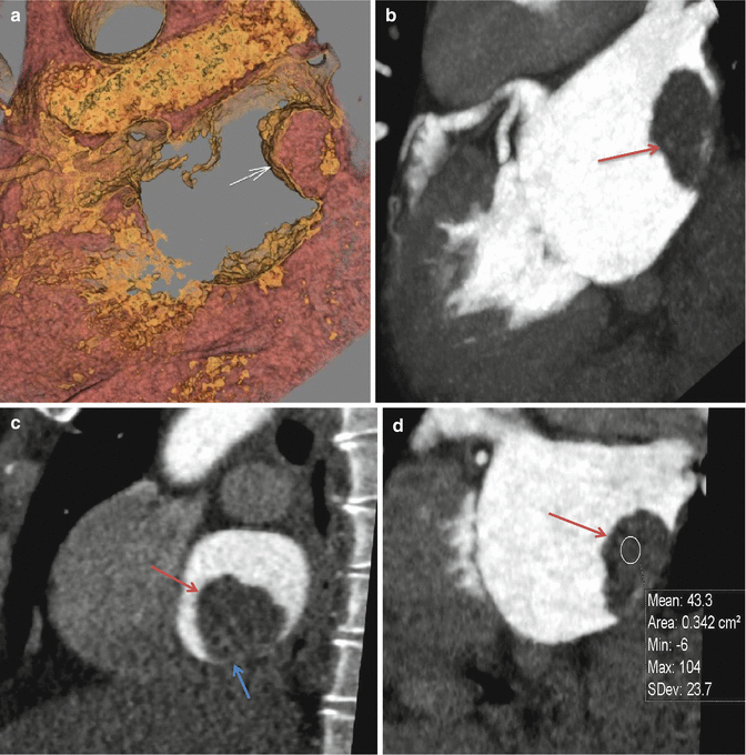
Myxomas are the commonest primary cardiac tumor. Although most often found in the atria, with a predilection for the left atrium, a myxoma may be present in any chamber [5, 6, 9–12]. They range in size from under 1 cm and can grow up to 10 cm [13] and may be sessile or pedunculated [5]. Pedunculated myxomas, if large enough, may be pushed into the orifice of the mitral valve during systole resulting in outlet obstruction (this is body position dependent) [5, 13]. Over time, this may cause damage to the mitral valve leaflets. Systemic embolization is a major complication of left-sided atrial myxomas and should be removed once discovered. Myxomas can occur in isolation or in a constellation of multiple additional cardiac and extracardiac findings, including cardiac and extracardiac myxomas, dark skin spots, and hyperactivity of the endocrine system [5, 14]. This is known as Carney syndrome and demonstrates autosomal dominant transmission [5]. Echocardiography is typically the initial imaging modality and demonstrates an echogenic mass within the cardiac chambers [15–17]. CT will demonstrate a soft tissue attenuation mass within the cardiac chamber with variable, heterogeneous enhancement and internal vascularity [15–17]. A stalk may be demonstrated in pedunculated myxomas. This is differentiated from an intracavitary thrombus based on its heterogeneous enhancement and internal vascularity (thrombi will not enhance and will not have internal vascularity) [15–17]. It is also differentiated from a lipoma based on its soft tissue attenuation (lipoma will have a fatty attenuation). On MRI, myxomas are typically hypo- to isointense on T1 and hyperintense on T2 [15–17].
7.2 Fibroelastoma
Papillary fibroelastomas are the second most common benign cardiac tumors [18, 19]. They are morphologically bizarre tumors, containing many “frond” like projections emanating from a central stalk [18, 19]. They vary in size from 2 to 70 mm, with an average of approximately 9 mm [18, 19]. They are most often found on the valves of the heart, with predilection to the left, with involvement of the aortic valve in 36 % of the time and the mitral valve 29 % of the time [18, 19]. Thirty percent of patients with fibroelastomas are asymptomatic, found incidentally on autopsy or imaging for unrelated reasons [18, 19]. When symptomatic, patients present with symptoms of thromboembolic disease secondary to embolization of small pieces of the projections which break off or embolization of associated thrombus [18, 19]. Most common symptoms are stroke or TIA. Other symptoms may include myocardial infarction and PE [18, 19]. Although typically visualized by echocardiography, MR is gaining popularity as the modality of choice for evaluation of fibroelastomas [19–21]. On echo, they typically appear as pedunculated echogenic valvular or paravalvular mass [19–21]. On MRI, they are typically hypointense on T2-weighted sequences due to their high fibrous content and are best imaged on bright blood SSFP (steady state free precession) sequences [19–21].
7.3 Cardiac Lipoma
Cardiac lipomas are common benign tumor of the heart and are composed predominantly of mature fat cells [22–25]. They may occur in the subendocardium [5] and protrude inwards into the chamber lumen (most commonly located in the right atrium and the left ventricle [22–25]), in the epicardial fat, or within the myocardium itself [22–25]. They may also occur on valves [26, 27]. When patients are symptomatic, the symptoms depend on the location of the lipoma [28]. When located within a cardiac chamber, they may result in outlet obstruction secondary to a ball in valve mechanism at the chamber outlet orifice [28]. This may also lead to valvular damage and insufficiency over time [28]. When present within the myocardium, the patient may present with conduction abnormalities and arrhythmias [28]. Cardiac lipomas may be imaged using echocardiography, CT, or MRI. On CT, they are typically well-circumscribed masses which demonstrate a fatty attenuation (HU <20), and on MRI, these lesions follow fat signal on all sequences and will drop their signal on fat suppression [22–25]. They are encapsulated by a fibrous capsule which appears hypointense on T1-weighted sequences [22–25]. They typically do not enhance on CT or MRI. Lipomatous hypertrophy of the intraatrial septum is not believed to be a true tumor, but rather brown fat that is trapped in the septum that has failed to resorb [22–25]. Sparing the fossa ovalis, they typically have a bilobed or dumbbell shape and lack a surrounding fibrous capsule [22–25].
7.4 Primary Malignant Cardiac Tumors
As mentioned above, malignant cardiac tumors are usually secondary to metastatic disease from a known primary neoplasm elsewhere in the body. Of the malignant primary cardiac tumors, the most common are sarcomas such as angiosarcoma and rhabdomyosarcoma, leiomyosarcomas, liposarcomas, and fibrosarcoma [6, 29–32].
7.5 Primary Cardiac Lymphoma
Cardiac lymphoma is often a component of diffuse lymphomatous metastatic disease and rarely occurs as a primary malignancy [33], accounting for 1.3 % of primary cardiac tumors and 0.5 % of extranodal lymphoma [33]. Cardiac lymphoma may involve the pericardium, epicardial fat, and myocardium. Pericardial thickening with pericardial effusion are early signs [33]. They often appear as ill-defined, infiltrative masses of equal to slightly lower attenuation than myocardium [33] and most commonly affects the right atrium [33]. Cardiac lymphoma demonstrates variable signal and enhancement characteristics on MRI [15, 33]. Clinical presentation of cardiac lymphoma is also variable, including pericardial effusion, arrhythmias, and nonspecific ECG abnormalities, most often a third-degree AV block [33].


Fig. 7.1
(a) A two-chamber hollow volume rendered image demonstrates a large mass adhered to the left atrial wall (white arrow). (b) Maximum intensity projection near two-chamber image demonstrates a well-circumscribed, lobulated, pedunculated mass within the left atrium which extends from the inferior atrial wall in a sessile configuration (red arrow). (c) An oblique-sagittal MPR view demonstrate the mild heterogeneous enhancement at the base (blue arrow). (d) Coronal reconstructed image demonstrates the mass to have a soft tissue attenuation of 43.3 HU. These findings are diagnostic of left atrial myxoma
< div class='tao-gold-member'>
Only gold members can continue reading. Log In or Register to continue
Stay updated, free articles. Join our Telegram channel

Full access? Get Clinical Tree


