24 Cardiac Tumor: Rhabdomyoma
I. CASE
A. Fetal echocardiography findings
a. The fetal echo reveals situs solitus of the atria, levocardia, left aortic arch, and heart rate of 150 bpm.
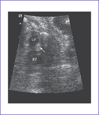
b. The four-chamber view is abnormal, with normal-sized ventricles each with echogenic masses in the walls.
c. The cardiac axis, position, and size are normal (cardiothoracic ratio = 0.3).
d. There is no evidence of compromise of the cardiac flows by color Doppler examination, but there is localized decreased left ventricle (LV) wall motion near one of the masses (Fig. 24-1A).
e. The inferior vena cava (IVC) is not dilated and has a normal blood flow pattern and velocity.
f. The systolic ventricular function of the right ventricle (RV) is normal.
g. There is no ascites or truncal edema.
h. Umbilical venous and arterial flow patterns are normal.
i. The aortic arch is leftward, and the ductal and aortic arches are of similar size. Both arches have antegrade flow.
j. Flow through the foramen ovale is unrestricted with a bidirectional shunt.
k. The pulmonary venous flow pattern is normal.
l. RV and LV Tei indices (myocardial performance index) are normal.
m. The ventricular walls show many small echogenic foci consistent with multiple tumors.
a. Similar intracardiac anatomy as twin A is seen but without congenital heart disease.
b. The RV apex contains a spherical echogenic tumor typical of rhabdomyoma (see Fig. 24-1B).
D. Fetal management and counseling
1. Amniocentesis was not offered because the risk from amniocentesis is probably higher than the risk of abnormal karyotype in cardiac tumor.
2. Follow-up included serial antenatal studies at 4-week intervals.
c. Arrhythmias such as ventricular or atrial ectopy or tachycardia can result from a focus near a tumor.
d. Imaging of the fetal brain could reveal subependymal nodules as well as cortical tumors (diagnosed by MRI) suggesting tuberous sclerosis complex (TSC).
F. Neonatal management
a. Evaluation should include standard vital signs, pulse oximeter, and blood pressures.
b. An electrocardiogram (ECG) is obtained to evaluate for arrhythmias or signs of myocardial ischemia.
c. An echocardiogram is immediately performed to assess ventricular function and outflow tracts.
a. Surgery is only indicated when there is critical inflow or outflow obstruction by the tumor, resulting in hemodynamic compromise with or without the need for ductal patency. In such a situation, the tumor should be resected.
b. Arrhythmia with incessant tachycardia and compromised hemodynamics could be managed with surgical resection, but with mapping of which tumor is causing the abnormal rhythm. However, with multiple cardiac tumors, it may be impossible to detect the source of the arrhythmia.


