Management
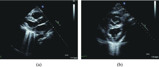
Right and left chest tubes were inserted into the respective pleural spaces. Approximately 1500–1800 cc of colorless fluid was evacuated from each pleural cavity. The patient’s ventilation improved dramatically, allowing us to perform TTE and transesophageal echocardiography that did not show a significant pericardial effusion. A small amount of fluid was present anterior to the RA (Figure 96.2, Videoclip 96.2).
Figure 96.2 After approximately 1500–1800 cc of colorless fluid was evacuated from each pleural cavity (a and b), there was no signs of pericardial effusion.
Discussion
Stay updated, free articles. Join our Telegram channel

Full access? Get Clinical Tree


