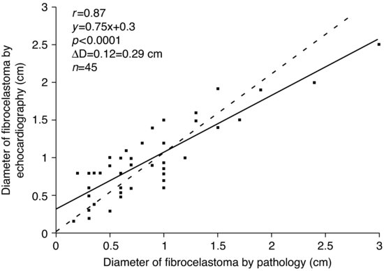
Figure 62.2 Comparison of tumor size measured by pathology and echocardiography in a subgroup of 45 patients shows a good correlation between two methods. Reproduced with permission from Elsevier.

The masses in the cardiac chambers were larger than those on the aortic or mitral valves (12.0 ± 4.6 mm versus 8.5 ± 4.4 mm in diameter, respectively; P < 0.001). All 19 CPFs in the chambers were mobile, as were 29 of the 91 on a valvular surface (31.9%). Single lesions were detected by echocardiography in 85 patients (91.4%). Multiple CPFs (range, 2–8) were detected in eight patients (8.6%). One patient had eight tumors observed in various locations on the right and left sides of the heart.
Echocardiographic follow-up data was available for 64 of 141 patients (45.4%) after surgical excision. The average follow-up time was 630 ± 903 days. No mass was detected by echocardiography in any patient during the follow-up period.
Discussion
Stay updated, free articles. Join our Telegram channel

Full access? Get Clinical Tree


