Cardiac Myxoma


Cardiac Myxoma


Joseph J. Maleszewski, M.D.

Allen P. Burke, M.D.


Definition and Histogenesis

Cardiac myxoma is a benign neoplasm characterized by stellate, ovoid, or plump spindle cells (so-called myxoma cells) embedded in a vascular myxoid stroma.1 While myxoid tumors occur with some frequency in the heart, cardiac myxomas are distinct neoplasms with characteristic histopathologic findings.

While initially thought to represent a reactive phenomenon, heterotopic tissue rests, or organizing thrombi, cardiac myxomas are now considered a neoplastic process by virtue of their location, characteristic histopathology, protein expression, and genetics.2,3,4 The precise etiology of the neoplastic cell, the “myxoma” (or lepidic) cell, is largely unknown. The diversity of cell types found in myxomas, such as endothelial cells, glandular elements, and apparently undifferentiated cells, has led many investigators to conclude that myxomas are of primitive multipotential mesenchymal origin.5,6


Epidemiology

Cardiac myxoma is the most common primary neoplasm of the heart. While other tumors (e.g., papillary fibroelastoma [PFE]) may occur at higher frequency, they do not appear to represent neoplastic processes.7 They can occur at any age, but most present in the fourth to sixth decades of life. In individuals <65 years of age, there is a female predilection with a male to female ratio of ˜1:2.8 In older individuals, the ratio is much closer to 1:1.9,10


Clinical and Radiologic Features


The Carney Complex

Approximately 3% to 10% of cardiac myxomas occur in the setting of the Carney complex (CNC; also known as the myxoma syndrome).4 This autosomal dominant condition is characterized by the constellation of myxomas (cardiac or otherwise), endocrinopathy (Cushing syndrome and/or acromegaly), and spotty skin pigmentation (particularly of the vermillion border of the lips). Diagnosis rests on recognition of major criteria or a combination of major and supplemental criteria in a given patient (Table 181.2).13

Aside from clinical criteria, inactivating mutations in the PRKAR1A gene have been found to be associated with CNC. Pathogenic PRKAR1A mutations have been found in more than 60% of individuals with CNC.13 Genetic testing for such mutations is available clinically and can help to identify and screen CNC kindreds.

Cardiac myxomas arising in the setting of CNC are often multiple and found in atypical locations (outside of the left atrium). Additionally, they are more likely to occur in younger individuals (average age of 25 years) and recur throughout life.


Imaging

Chest radiography may show cardiomegaly and/or pleural effusions.14 Calcification may be seen in up to 30% of cases and seems to be slightly more common in right atrial myxomas.9,14 Echocardiography is a sensitive test of cardiac masses, including myxoma. Computed tomography (CT) and cardiac magnetic resonance imaging are often performed for preoperative characterization of tissue density and documentation of the relationship of the tumor to adjacent structures.15 On CT, there is often a heterogeneous signal of the endocardium-based lesion.16


Prognosis

Despite the benign nature of the neoplasm, like other benign cardiac tumors, myxomas can rarely result in devastating consequences, depending on their size, location, and gross characteristics. Villous tumors and tumors containing >50% myxomatous stroma are more likely to embolize, and large tumors are more likely to obstruct. Either of these phenomena could result in sudden death.

Following recognition of the tumor, the prognosis of cardiac myxomas is largely driven by whether or not the tumor is arising in a
syndromic context. Those tumors occurring in the setting of CNC have a relatively high rate of recurrence (10% to 20%), while those outside of CNC have a very low recurrence rate (<5%).17,18 Most recurrences occurring in nonsyndromic tumors occur as a result of incomplete tumor resection, which is why it is recommended that the cardiac surgeon take a rim of normal tissue at the attachment site to ensure complete excision.


TABLE 181.2 Diagnostic Criteria for Carney Complexa


Major criteria


Cardiac myxoma


Other myxoma (e.g., cutaneous, mucosal, breast)


Lentigines (particularly at the vermillion border)


Cushing syndrome


Acromegaly


Large cell calcifying Sertoli cell tumor


Psammomatous melanotic schwannoma


Osteochondromyxoma


Supplemental criteria


Affected first-degree relative


Inactivating mutation of the PRKAR1A gene


aDiagnosis requires 2 major criteria or 1 major + 1 supplemental criterion.


Adapted from Stratakis CA, Kirschner LS, Carney JA. Clinical and molecular features of the Carney complex: diagnostic criteria and recommendations for patient evaluation. J Clin Endocrinol Metab. 2001;86:4041-4046.


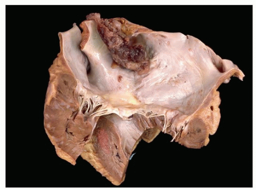
FIGURE 181.1 ▲ Left atrial cardiac myxoma arising from the septum. The sizeable tumor is protruding into the left atrial cavity and partially obstructed blood flow through the mitral valve.


Pathologic Features


Location and Gross Pathology

Cardiac myxomas are endocardial lesions that protrude into the cardiac chamber from which they arise. Most (80% to 90%) arise in the left atrium, with attachment to the atrial septum in the region of the fossa ovalis (Fig. 181.1). As benign neoplasms, cardiac myxomas do not (by definition) invade, a point that serves to differentiate these lesions from malignant cardiac sarcomas or metastatic lesions. While these tumors have been described in all cardiac chambers, the finding of a cardiac myxoma outside of the left atrium should raise concern for the possibility of a CNC-associated lesion. Valvular myxomas have been reported, but are distinctly unusual.


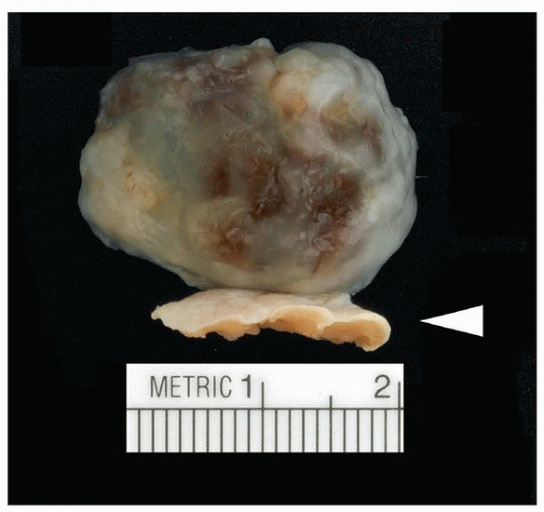
FIGURE 181.2 ▲ Cardiac myxoma with endocardial attachment (arrowhead). A rim of the myocardium is typically taken with the tumor at surgical resection to ensure completeness of excision.


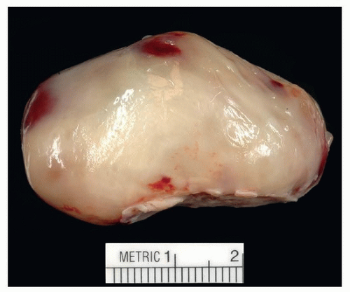
FIGURE 181.3 ▲ A smooth surface tumor with a relatively broad base (bottom).

The tumors range in size from only a few millimeters to more than 15 cm. They may either arise from a stalk or be sessile and broad based (Fig. 181.2). Grossly, they can be either smooth surfaced or villous (Figs. 181.3 and 181.4). The latter appear to be more associated with
thromboembolic symptoms, either from adherent surface thrombus on the finger-like projections of tumor or from the friable tumor itself.19

Only gold members can continue reading. Log In or Register to continue

Stay updated, free articles. Join our Telegram channel

Aug 19, 2016 | Posted by in CARDIOLOGY | Comments Off on Cardiac Myxoma

Full access? Get Clinical Tree

Get Clinical Tree app for offline access