CHAPTER 6 Cardiac masses, infection, congenital abnormalities
6.1 CARDIAC MASSES
Echo is very important in detecting cardiac masses and giving an indication of their nature. Masses include:
1. Tumours of the heart
Primary tumours – rare
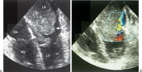
Echo cannot differentiate between benign and malignant tumours. 2-D echo shows tumours as echogenic masses within the cavity of the heart, attached to the wall or in the pericardium. The size and mobility can be determined. As with all echo studies, multiple views should be obtained. Occasionally on M-mode, a tumour such as myxoma may be seen interfering with valve function (section 2.1). The effects of tumours (e.g. obstruction of valve flow, LV dysfunction due to infiltration or obstruction or pericardial effusion) can also be seen on echo (Fig. 6.1).
Myxoma
The effects of myxomas relate to:
Myxomas usually present in one of four ways, in decreasing order of frequency:
Myxomas may be readily detected by M-mode or 2-D echo (Fig. 6.2). The myxoma can be seen as a mass in the LA cavity and may prolapse through the MV into the LV cavity during diastole obstructing flow. It may be so large as to fill the LA. Doppler can show the haemodynamic effects.
Myxomas very rarely occur in an autosomal dominant familial fashion associated with lentiginosis (multiple freckles) or HCM and so it is wise to screen all first-degree relatives by echo (section 7.6).
2. Thrombus
Some examples of these situations include:
False-positive identification of thrombus may occur due to:
The following favour the diagnosis of thrombus:
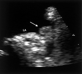
A number of echo views should always be taken. On 2-D echo, thrombus may be seen as a ball-like or a frond-like mass, or as a well-organized, laminated, raised thickening in the LA or LV. In the LA, there may be associated evidence of sluggish blood flow such as ‘spontaneous contrast’. The LA appendage may contain thrombus which can be identified on TOE (Fig. 6.3).
6.2 INFECTION
Endocarditis
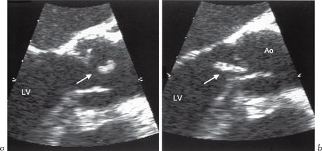
This refers to inflammation of any part of the inner layer of the heart, the endocardium, including the heart valves. Inflammatory and/or infected material may accumulate on valves to cause masses called ‘vegetations’. These are made up of a mixture of infective material, thrombus, fibrin and red and white blood cells. Vegetations are usually attached on valves but may be on other locations, e.g. chordae, LA, LVOT (HCM), right side of VSD (jet lesion).
The size of vegetations varies from <1 mm to several cm. TTE can miss vegetations <2 mm. TOE may show these, and improves sensitivity to >85%. Large vegetations are particularly associated with fungal infection or endocarditis of the tricuspid valve. Vegetations may be detected by M-mode (section 2.1) or 2-D techniques where they are seen as mobile echo-reflective masses.
There are a number of potential causes which may be infective or non-infective.
Infective endocarditis
Endocarditis is a serious condition and potentially life-threatening. It can be acute (e.g. with Staphylococcus aureus) or subacute bacterial (SBE). Infection usually follows an episode of bacteraemia, which may not be readily identifiable, or may follow dental treatment or surgery. For this reason it is safest to advise antibiotic prophylaxis treatment for all dental treatment and all surgical procedures for individuals with a known cardiac murmur, congenital lesion, heart valve abnormality or artificial valve. Certain infecting organisms are associated with underlying disease conditions, e.g. Streptococcus bovis endocarditis with carcinoma of the colon.
Clinical features supporting endocarditis
Important investigations in endocarditis
Cardiac lesions predisposing to endocarditis
1. Common
Antibiotic prophylaxis to prevent endocarditis
The recommended antibiotic regimen should be checked locally and must include attention to a subject’s known antibiotic allergies. Used in:


