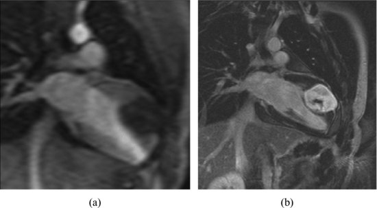
The magnetic resonance images showed a regular mass in the myocardium of LV lateral wall close to the apex (Figure 63.2).
Figure 63.2 The magnetic resonance images showed a regular mass in the myocardium of the lateral free wall of the left ventricle close to the apex (a). Magnetic resonance images postgadolinium enhancement (b).
Treatment
The patient was referred to cardiothoracic surgery for complete resection of a benign neoplasm. After operation, he resumed normal sports activities.
Surgical/pathologic findings: Cardiac fibroma measuring 8 cm × 6 cm × 6 cm was found. It was completely encapsulated and was with irregular margins.
Discussion
Stay updated, free articles. Join our Telegram channel

Full access? Get Clinical Tree


