Cardiac Computed Tomography and Magnetic Resonance Imaging

8 Cardiac Computed Tomography and Magnetic Resonance Imaging


The past decade has seen rapid development in cardiovascular imaging technologies coupled with novel clinical applications. Noninvasive imaging technologies now allow for assessment of cardiac morphology, function, perfusion, and metabolism. The explosion in imaging has led to increasing financial expenditures. From 1999 to 2003, diagnostic imaging services reimbursed under the U.S. Medicare physician fee schedule grew more rapidly than any other type of physician service. Both cardiac computed tomography (CCT) and cardiac magnetic resonance imaging (CMR) have interesting and unique advantages compared with alternate imaging modalities. Understanding the applications and limitations of these modalities will permit optimal and efficient use in the future.


Cardiac Computed Tomography


Chest pain is a common clinical problem and one of the most common complaints of individuals presenting for urgent medical evaluation. One of the most important, life-threatening causes of chest pain is coronary artery disease (CAD). Although cardiac catheterization is the best method to assess for the presence of hemodynamically significant CAD available today, it is impractical as a screening test. It is invasive and costly, can be especially dangerous in some subsets of patients, and when used broadly as a screening tool is performed on a substantial number of patients who have no significant CAD and/or whose chest pain is unrelated to cardiac causes.


For decades, investigators have sought to develop new technologies that would allow rapid noninvasive imaging of the coronary arteries and other cardiac structures. One such technology that has evolved rapidly is CCT. Although not yet as accurate as cardiac catheterization, CCT now permits visualization of the coronary arteries and coronary lumen as well as assessment of cardiac function, the pericardium, left atrial anatomy, congenital heart disease, pulmonary arterial and venous anatomies, and diseases of the aorta.


Technology of CCT


Imaging the heart and coronary arteries with CT is challenging for several reasons and requires more sophisticated imaging and analysis approaches than are required for other body regions. Major difficulties arise because coronary arteries are relatively small structures with branches of interest in the range of 2 to 4 mm in diameter, and they are moving structures. The coronary arteries show rapid cyclic motion throughout the cardiac cycle—essentially moving in three dimensions with each heartbeat. Furthermore, when the subject breathes, the heart and vessels move within the chest. However, several major advances in recent years have dramatically improved the resolution of coronary artery images. These include acquiring more data/images at one time, decreasing the time patients must hold their breath; development of smaller CT x-ray detectors, increasing spatial resolution to visualize smaller structures; the development of scanners with increased rotational speed resulting in increased temporal resolution so that moving objects such as the arteries can be “frozen”; and the ability to gate the CT acquisition to the patient’s ECG, allowing visual reconstruction of the heart and arteries during different phases of the cardiac cycle. Typically, the coronary arteries have less motion in diastole when the heart is filling compared with systole when the heart is contracting. Temporal resolution is directly related to x-ray tube gantry rotation time. A standard single source (one x-ray tube) allows for a temporal resolution of 167 ms. Newer scanners from some vendors afford dual-source CT technology (two x-ray tubes in the gantry), resulting in an effective scan time of 83 ms independent of heart rate. The small size of cardiac structures requires excellent spatial resolution, which is on the order of 0.4 to 0.75 mm with current technology. Respiratory motion artifact is minimized by asking the patient to hold his or her breath during image acquisition. Even with these advances, CCT lacks the resolution attained in the cardiac catheterization laboratory where images can be obtained at 30 frames per second, yielding temporal resolutions that can be greater than 33 ms with spatial resolution less than 0.1 mm.


Several different CT technologies have been used for cardiac imaging. Electron beam CT (EBCT), initially introduced in the mid-1970s, utilizes an electron source reflected onto a stationary tungsten target to generate x-rays, allowing for very rapid scan times. EBCT is well suited for cardiac imaging because of its high temporal resolution (50–100 ms) with an estimated slice thickness of 1.5 to 3 mm and the ability to scan the heart in a single breath hold. This technology was initially used to quantify coronary arterial vessel wall calcium volume and density—generating a patient-specific score—and it remains the primary use of EBCT. Coronary calcium scores are independent of other traditional cardiac risk factors in the prediction of cardiac events and, as such, can be considered an excellent biomarker for the presence of CAD and the risk of future cardiac events. Efforts to use EBCT technology to visualize the lumen of the coronary artery with the administration of intravenous contrast agents have thus far proven to be limited, in large part as a result of the very limited spatial resolution.


EBCT has been largely supplanted by newer, multidetector CT (MDCT) technology, which involves a mechanically rotated x-ray source and offers increasing spatial resolution. New generations of scanners permit the simultaneous acquisition of more data (“slices”). These advances have allowed for markedly increased spatial resolution and for complete acquisition of data during one breath hold. Coronary calcium scoring can also be performed using MDCT with results that are comparable to those obtained by EBCT. What MDCT offers, however, is sufficient spatial resolution to make coronary CT angiography (CTA) feasible. Proof-of-concept studies were initially performed using MDCT machines capable of obtaining four to eight slices per scan.


As technology has advanced, 64-slice (and higher) scanners are now available and allow acquisition of higher resolution images without the requirement for long breath holds or extremely slow heart rates. It is currently recommended that CTA be performed using a minimum of a 64-slice scanner. These scanners are now commonly available in many hospitals. With this type of scanner, 64 simultaneous anatomic slices are acquired, allowing a complete cardiac study to be performed with one breath hold, typically in 10 to 15 seconds. Because of the limited temporal resolution, a successful diagnostic scan on a conventional 64-slice scanner requires that the heart rate be steady and usually less than 60 to 65 bpm. Newer prototypes allow up to 320 anatomic slices to be simultaneously acquired. With a minimal slice thickness of 0.75 mm, an entire heart can be imaged in a single heartbeat. Even with 320-slice scanners, temporal resolution does not reach what can be obtained routinely in a cardiac catheterization laboratory, and images are better in patients with relatively low heart rates. To overcome the necessity of a slow heart rate, one vendor has placed two x-ray sources in the scanner (so-called dual source). This technology offers an improved temporal resolution even with heart rates approaching 100 bpm and greater.


Data Acquisition Techniques


For CTA using a single-source scanner, it is necessary to image with heart rates less than 65 bpm. Most commonly, an oral or intravenous β-blocker is given to slow the heart rate. In some settings, sublingual nitrates may be administered to dilate the coronary arteries and allow them to be more easily imaged. Coronary CTA requires intravenous administration of a contrast agent to opacify the lumen of the coronary arteries. The intravenous contrast agents used for CTA carry the same dose-dependent risks in patients with renal dysfunction as contrast agents used for cardiac catheterization, as well as the risk of an allergic reaction to iodine. Respiratory motion is minimized by patient breath hold from 6 to 20 seconds, depending on scanner generation and cardiac dimension. Data acquisition varies somewhat based on scanner type. The most common data acquisition protocol utilizes a spiral mode involving continuous data acquisition during constant rotation of the x-ray tube while the patient is simultaneously continually advanced on the table through the x-ray gantry. To minimize radiation exposure, data acquisitions can be performed in sequential mode (step and shoot). This involves acquisition of single transaxial slices sequentially as a patient is advanced stepwise through the scanner.


Excessive cardiac motion can lead to blurring of the contours of the coronary vessels. For this reason, a regular heart rate is necessary for optimal imaging of the coronary arteries. Relative contraindications to performing CTA include the presence of frequent ectopic beats or atrial fibrillation. Coordinating data acquisition and analysis to the cardiac cycle involves either prospective triggering or retrospective gating. In prospective triggering, data are acquired in late diastole, based on simultaneous ECG recordings. In retrospective gating, data are collected during the entire cardiac cycle. Post-processing then allows only data from specific periods of the cardiac cycle to be used for image reconstruction.


Clinical Indications


Coronary Artery Calcium Score


Coronary artery calcium (CAC) is recognized as a marker of subclinical atherosclerosis. CAC scoring utilizes no contrast and readily detects calcium because of its high x-ray attenuation coefficient (or CT number) measured in Hounsfield units (HU) (Fig. 8-1). The Agatston scoring system assigns a calcium score based on maximal CT number and the area of calcium deposits. Initially promoted as part of a screening paradigm, CAC was originally made available for patient-initiated evaluation of coronary risk on a fee-for-service basis. More recently, analysis of several large clinical datasets has confirmed that the “coronary calcium score” is a predictor of coronary events, independent of traditional risk factors. In at least one study, calcium score was more predictive than C-reactive protein and standard risk factors for predicting CAD events.
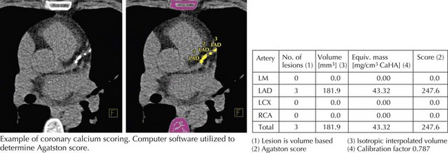


The coronary calcium score is derived by identifying coronary arterial tree segments that have attenuation characteristics (HU) greater than 130 that correlate with the attenuation due to calcium. These calcified lesions are scored by size and density with a weighting factor for increasing density. Technically, the score reflects analysis of contiguous pixels in the x, y, and z directions that are calcium-positive. Discrete lesions are scored separately, and the density of calcium within each lesion is graded from 1 to 4 according to the HU. The sums of all the lesions are totaled to arrive at a single coronary calcium score. In general, the higher the score, the greater the amount of calcified plaque within the arterial tree. There is a positive correlation of cardiac events with this score. Many individuals younger than 50 years have no coronary artery calcification and thus have a calcium score of 0.


The Multiethnic Study of Atherosclerosis (MESA) Group published a series of articles suggesting that the calcium score is an independent risk factor for cardiac events. Also, MESA’s website has the capacity to allow comparison of an individual patient’s calcium score against their large database. This score takes into account age, sex, and race, and generates a percentile compared to the database studies. The 2007 American College of Cardiology (ACC)/American Heart Association (AHA) Clinical Expert Consensus Document on CAC scoring states that in patients with intermediate coronary heart disease risk (10%–20% 10-year risk of estimated coronary events), it may be reasonable to consider use of CAC measurement based on evidence that it demonstrates incremental risk prediction such that patients might be reclassified to a higher risk status and subsequently initiated on pharmacotherapy, particularly for cholesterol lowering. The presence of a high calcium score may prompt clinicians to use more aggressive therapy as if they were reclassified in a higher risk group, or to convince patients who are reluctant to take drugs such as statins to take their disease more seriously.


CTA utilizes intravenous contrast to differentiate vessel lumen from vessel wall. In 2006, the ACC and many other societies with interests in cardiac imaging put together recommendations of “appropriateness criteria” for utilization of cardiac CTA that include appropriate (Box 8-1) and inappropriate uses of this technology. The most common appropriate utilization is diagnostic study of patients presenting with chest pain who do not have significant ECG changes or elevated cardiac biomarkers but have an intermediate probability of CAD. At experienced centers with careful data acquisition, sensitivities range from 83% to 99% and specificities from 93% to 98% with remarkably high estimated negative predictive value (95%–100%), indicating that CCT may be used to reliably rule out the presence of significant flow-limiting coronary atherosclerotic disease. It should be pointed out that CCT would be inappropriate for patients at high risk for or with other indications of cardiac ischemia such as elevated biomarkers or significant ECG changes. Those patients should be referred immediately for invasive imaging.


Bypass graft imaging is more easily accomplished than coronary artery imaging because of the larger size of bypass grafts (particularly saphenous vein grafts) and less rapid movement of bypass grafts as compared with native coronary arteries. The patency or occlusion of grafts can be determined by the presence or absence of distal target vessel contrast enhancement (Fig. 8-2

Only gold members can continue reading. Log In or Register to continue

Stay updated, free articles. Join our Telegram channel

Jun 12, 2016 | Posted by in CARDIOLOGY | Comments Off on Cardiac Computed Tomography and Magnetic Resonance Imaging

Full access? Get Clinical Tree

Get Clinical Tree app for offline access