(1)
University of Ottawa The Ottawa Hospital, Ottawa, ON, Canada
Sudden cardiac death accounts for >50 % of all coronary artery disease deaths and 15–20 % of all deaths (Gillum 1990; Myerburg et al. 2004). Thus cardiac arrest prevention represents a major opportunity to further reduce mortality (Deo and Albert 2012).
Survival to hospital discharge was estimated to be only 7.9 % among out-of-hospital (OOH) cardiac arrests that were treated by emergency medical services personnel (Nichol et al. 2008). Importantly, the majority of cardiac arrests occur at home, mostly unwitnessed. Thus, automated external defibrillators (AEDs) that improve resuscitation rates for witnessed arrests have limited effectiveness on reducing overall mortality. Should most homes with occupants older than age 50 deemed to be at risk carry a defibrillator?
Dr J.F. Pantridge initiated coronary care ambulances in Belfast in 1966 (Pantridge and Geddes 1967) and was instrumental in developing portable defibrillators. He advocated that a cardiac defibrillator should be considered in the same light as a fire extinguisher and thus should be available in most homes, place of work, and sports arenas (J.F. Pantridge, 1974 personal conversation).
“Time Is of the Essence”
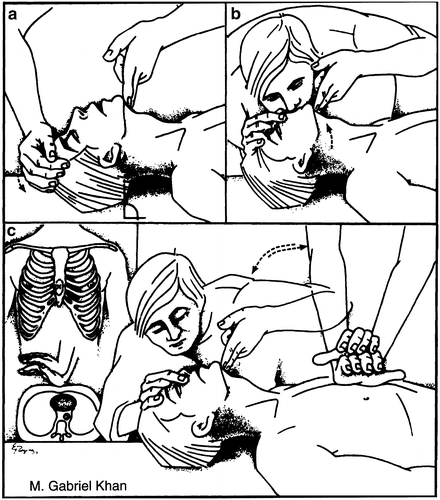
In the majority of cases of cardiac arrest, ventricular fibrillation (VF) is the only correctable rhythm. Thus, there is no need to waste precious time checking for a pulse or for the rhythm prior to defibrillation. Just get on with 100 compressions/min and continue for 4 min (Bray et al. 2011) while someone grabs a defibrillator and rapidly defibrillate. Only halt the 100/min compressions when the defibrillator is charged and ready to apply. See Fig. 15-1.


Fig. 15-1
Sequence is CAB, not the old ABC. Compress hard and fast, 100/min, until an AED is available for immediate defibrillation; then continue compressions until circulation restored. Following defibrillation, rescuers should not interrupt chest compressions to check circulation (e.g., evaluate rhythm or pulse) until after approximately 2 min of CPR. No need for rescue breath until after the fourth minute (in the United Kingdom 6 min.). Note the position and shape of the applied hands
A “dispatcher should provide instructions assertively on compression-only CPR. Thus the ‘kiss of life’ should be replaced by ‘Keep It Simple, Stupid’, which is broadly consistent with the practice of many emergency medical dispatchers in the UK” (Nolan and Soar 2010).
Following defibrillation, immediately continue compressions.
No need for rescue breath until after the fourth minute (in the United Kingdom 6 min).
Approx 50 % of patients with VF survive cardiopulmonary resuscitation (CPR) if a defibrillator is available, versus <10 % for other rhythms, represented by asystole and pulseless electrical activity. The incidence of VF in most surveys of cardiac arrest is approx 50 %. Thus chest compression and immediate defibrillation remains the mainstay of therapy for cardiac arrest.
Two Cardiac Rhythms
There are only two cardiac arrest rhythms:
1.
Ventricular fibrillation/pulseless ventricular tachycardia (VF/VT). VF is defined as a pulseless chaotic disorganized rhythm with an undulating irregular pattern that varies in size and shape and a ventricular waveform >150/min.
2.
Non-VF/VT: asystole and pulseless electrical activity.
Individuals who can be saved from cardiac arrest are usually in VF.
The widespread distribution of AEDs has helped to accomplish early defibrillation programs and has definitely saved lives particularly in crowded stadiums.
All first-responding emergency personnel, both hospital and nonhospital (e.g., physicians, nurses, emergency medical technicians, paramedics, firefighters, volunteer emergency personnel), must be trained and permitted to operate a defibrillator, which should be readily available in all emergency ambulances or emergency vehicles that engage in the transit of cardiac patients and should be available in many areas including shopping centers and sports arenas. Availability of the AED is thus of paramount importance.
Modern biphasic defibrillators have a high first-shock efficacy in more than 90 % (White et al. 2005; Morrison et al. 2005); thus, VF is usually eliminated with one shock of 360 J.
In a study of 168 consecutive cases of sudden coronary death (within 6 h of onset of symptoms), Davies (1992) showed that 74 % had a recent coronary thrombotic lesion. These patients had warning chest pain prior to arrest. In 52 patients without chest pain or infarction, 48 % had coronary thrombi. In patients with previous infarction, the absence of warning chest pain before arrest selects for a primary arrhythmia caused by preexisting hypertrophy or scarring or both. In a study of successfully resuscitated cardiac arrest victims, 40 of 84 (48 %) had coronary artery occlusion on angiography; successful angioplasty was achieved in 28 of 37 of these patients (Spaulading et al. 1997).
Atheroma complicated by erosion, fissuring, or minute rupture initiates thrombus formation and cardiac arrest; thus the author recommends daily aspirin 75–81 mg non-enteric coated to be taken after the evening meal to all who are at risk, typically:
Men over age 45 with family history of MI prior to age 75 (old concept < age 55)
Cigarette smokers
Patients with mild to moderate hypertension
Sedentary men > age 45
Male > age 40 and female > age 50 with LDL cholesterol > 3.5 mmol/l ~ 140 mg/dl
Male > age 45 and female > age 50 with stressful occupation
Male > age 45 and female > age 50 who spend > 1 h/day in cities driving in high-volume traffic and cannot avoid pollution from automobile exhaust fumes (see Chap. 11 for details)
Diabetics > age 40
Hopefully the antithrombotic effects of aspirin may prevent enlargement of thrombus, by ameliorating platelet aggregation. This scenario will never be tested in an RCT but is logical therapy. Individuals with known peptic disease or GI bleeding should not consider this strategy.
Cardiac deaths in young athletes are usually caused by hypertrophic cardiomyopathy (Maron 1997) (35 %) and anomalous origin of the left main coronary artery from the right sinus of Valsalva (20 %). Other conditions causing sudden cardiac death include arrhythmogenic right ventricular dysplasia, prolonged QT syndromes, and Brugada syndrome (See Chap. 14).
When multiple rescuers are present, one rescuer can perform compression CPR while the other readies the defibrillator, thereby providing both immediate compressions and early defibrillation.
A change from a three-shock sequence to one shock followed immediately by compressions.
The consensus recommendation for initial and subsequent monophasic waveform doses is 360 J.
Modern biphasic defibrillators have a high first-shock efficacy (defined as termination of VF for at least 5 s after the shock), averaging more than 90 %, so that VF is likely to be eliminated with one shock.
Basic Life Support
Adult basic life support (BLS) (AHA Guidelines 2010a, b, 2012) requires:
Rapid recognition of cardiac arrest (assessing responsiveness and the absence of normal breathing); victims of cardiac arrest may initially have gasping respirations or even appear to be having a seizure. The lay rescuer should not attempt to check for a pulse and should assume that cardiac arrest is present if an adult suddenly collapses, is unresponsive, and is not breathing or not breathing normally (i.e., only gasping).
Activation of the emergency response system.
Rapid commencement of high-quality chest compression.
See Fig. 15-1. Place the heel of one hand over the lower half of the sternum but at least 1½ in. (two fingerbreadths) away from the base of the xiphoid process. The heel of the second hand is positioned on the dorsum of the first, both heels being parallel. The fingers are kept off the rib cage; if the hands are applied too high, ineffective chest compression may result, and fractured ribs are more common with this hand position:
Position of the hands on each other is crucial to obtain downward pressure.
No bend at the elbows.
Hand on the chest avoiding the rib cage (see Fig. 15. 1).
THE bystander or trained person should push hard and fast on the patient’s chest with the goal of compressing at a rate of at least 100 times/min at a depth of at least 2 in. (AHA Scientific Statement 2012).
Rapid defibrillation.
The recommended depth of compression for adult victims has increased from a depth of 1–2 in. to a depth of at least 2 in.
The arms are kept straight at the elbow (locked elbows), and pressure is applied as vertically as possible. The resuscitator’s shoulders should be directly above the victim’s sternum. The rescuer should compress at the center of the chest at the nipple line.
Chest compressions are straight down and thus easily carried out by forceful movements of the shoulders and back, so the maneuver is less tiring. The sternum is depressed 2 in. toward the spine using the heel of both hands. CPR should never be interrupted for more than 10 s. This interval should be sufficient to allow defibrillation. Cessation should not exceed 30 s for endotracheal intubation.
The 2010 AHA Guidelines for CPR is a change in the BLS sequence of steps from “A-B-C” (airway, breathing, chest compressions) to “C-A-B” (chest compressions, airway, breathing) for adults (see studies : Svensson et al. 2010, Rea et al. 2010a).
Studies indicate that an AED must be available within 5 min of the arrest to engage the shockable rhythm or the result is a poor outcome. In the majority of cases of cardiac arrest, when an AED is available somewhere in the facility, it usually takes >5 min to obtain the AED at the side of the victim, and thus bystanders who witness the arrest must be encouraged to learn and initiate chest compressions within seconds of the witnessed patient collapse and to give 100 compressions/min until the defibrillator is in readiness to apply a shock (4–6 min).
< div class='tao-gold-member'>
Only gold members can continue reading. Log In or Register to continue
Stay updated, free articles. Join our Telegram channel

Full access? Get Clinical Tree


