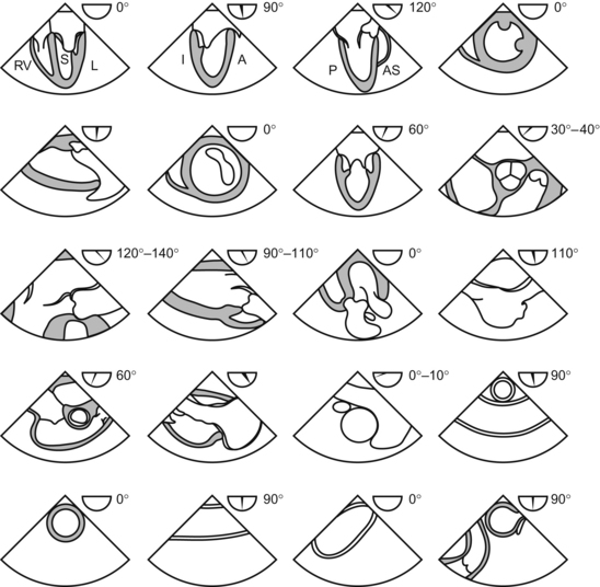
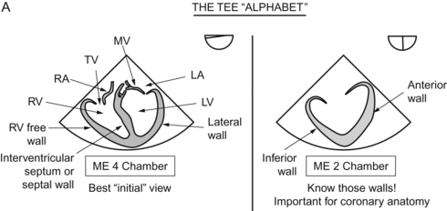
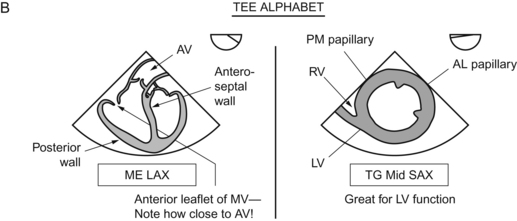
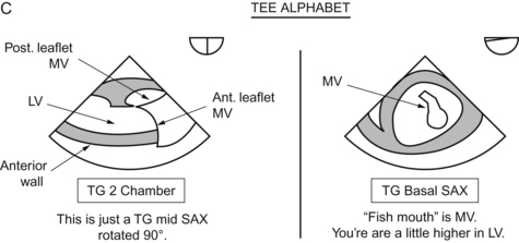
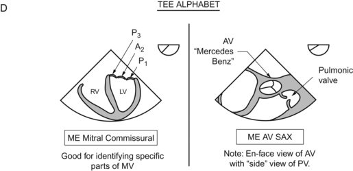
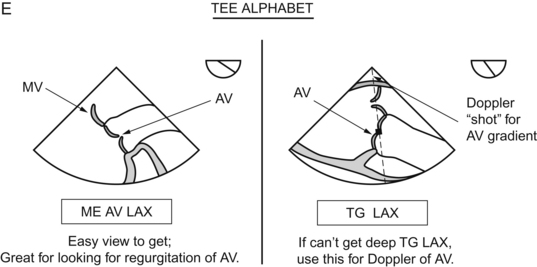
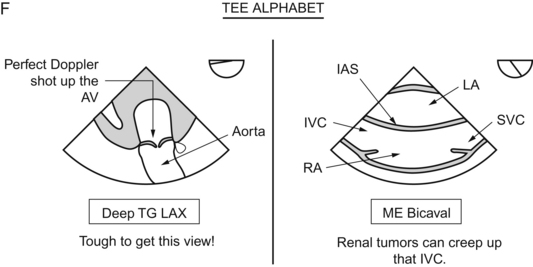
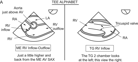
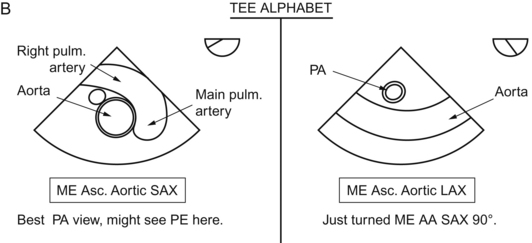
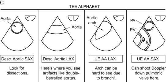
Chapter 9 Cardiac Anatomy Christopher J. Gallagher and John C. Sciarra Time to go back to the model and the pie-shaped slice of imaging that comes out of the echo probe. And time to take another good look at a plastic model of a heart. These imaging planes tie in with the BIG 20 views of the heart. These are the 20 views detailed in THE ARTICLE by Shanewise et al on echo.1 The 20 views are also detailed a million times over on different internet sites. Google “University of Toronto: TEE” for a great tutorial. This article and the 20 views detailed therein are the absolute crux of the TEE experience. Photocopy those 20 views and tape them to your TEE machine. Every time you examine a patient, try to get all the 20 views. Get in the habit of “examining everything every time”. If not, you’ll just look at the “thing of interest” and you’ll miss something else. Shanewise has thrown down the gauntlet. Can you do it that fast? In each view, note the approximate omniplane angle! “In the bicaval view, I see an ASD.” “In the 4-chamber view, I see a clot in the apex.” “The ME 2-chamber view shows a new regional wall motion abnormality; the inferior wall is not moving.”
Imaging Planes
Thoracic Key
Fastest Thoracic Insight Engine


