Cardiac Amyloidosis


Cardiac Amyloidosis


Joseph J. Maleszewski, M.D.

Allen P. Burke, M.D.


Introduction


General

Amyloidosis is the deposition of misfolded protein with an antiparallel β-pleated sheet configuration in a variety of organs including the heart.1 Amyloidosis is generally systemic, although localized forms involving particular types of amyloid have been described, including in both the heart and lung (Chapter 27). Although the histologic appearance and histochemical staining characteristics are identical regardless of the protein precursor, amyloid typing has become essential, as treatment is directed at the underlying disease process or, in some instances, the protein itself.


Classification

The classification of the amyloidoses is evolving, but currently is driven primarily by the type of misfolded protein present in tissue (Table 148.1). Cardiac amyloidosis can be broadly divided into acquired and hereditary forms, the latter characterized by the deposition of mutant proteins. While light chain (AL)-type amyloidosis is responsible for most (˜80%) cases of cardiac amyloidosis overall, the majority (approximately two-thirds) of cases seen at cardiac biopsy for symptomatic amyloidosis are of the transthyretin (ATTR) variety, owing to selection biopsy of those that undergo endomyocardial sampling.

All amyloidoses are named by designating the abbreviated name of the protein with the letter “A” (meaning amyloid).2 Therefore, transthyretin-type amyloidosis is abbreviated ATTR, (A [amyloid] +TTR [the abbreviated name of the protein]). ATTR-type amyloidosis includes both mutant (hereditary) and acquired (wild-type) forms. The latter instance is sometimes referred to as senile systemic amyloidosis; however, the term wild-type ATTR amyloidosis is now strongly preferred. Alternatively, wild-type and mutant ATTR have been designated ATTRwt and ATTRm, respectively. Variants such as the V122I polymorphism can also be designated by indicating the variation after the amyloid type (e.g., ATTR V122I).3

Light chain amyloid is designated AL-type amyloidosis and is the result of an underlying plasma cell dyscrasia. Although amyloid derived from serum amyloid A protein (AA) is a relatively common form of systemic amyloidosis, it rarely if ever causes cardiac symptoms.

By far the most frequent hereditary amyloidosis to involve the heart is mutant or hereditary ATTR.

So-called isolated atrial amyloidosis, caused by deposition of atrial natriuretic factor (AANF), is characterized by relatively sparse amyloid depositions within the atrial walls. Such deposits usually occur in older individuals, though its relationship with disease has not yet been established.


Methods of Typing Amyloid

It has been shown that the presence of a monoclonal gammopathy is not adequate for typing of amyloid, because 15% of patients with AL-type amyloidosis have no evidence of such in their serum or urine
(“nonsecretors”). Furthermore, up to 25% of patients with ATTR-type amyloidosis have monoclonal immunoglobulins in their serum or urine.4,5 Because treatment is driven by the amyloid protein present, it is necessary to type amyloid when it is identified in cardiac biopsy samples.


TABLE 148.1 Cardiac Amyloidosis, Clinical Findings by Protein Precursor


Type


Amyloid Subunit


Age (mean)


Cardiac Involvement


Frequency of Patients Diagnosed with Cardiac Amyloid by Biopsy


Extracardiac Involvement


ATTR (senile systemic amyloidosis)


Trans-thyretin, wild typea


70-83


Invariable, usually mild but may be severe


30% (overall 26%-57%)


Predominantly in vessels, especially lung


ATTR (hereditary)


Trans-thyretin, mutant


57


Usual, especially severe with Leu55Pro, Val30Met, Val122Ile, Tyr78Phe mutations


12%-25%


Nerves, endocrine tissues, various viscera with sparing of liver and relative sparing of kidney


AL


Immunoglobulin light chains


56-60


Frequent


44% (λ) 43% all (M)


9% (κ)


Range 45-82


Multiorgan involvement, frequent involvement of liver, spleen, and kidney


Apo A1


Mutant apolipoprotein A1


Few cases


May be severe


<1


Skin


Kidney


Stomach


Larynx


Bowel


Liver


Gynecologic tract


Lymph nodes


AANF


Atrial natriuretic peptide


80+


Invariable (atria)


0


None


AA


Amyloid protein A


Frequent, rarely symptomatic


0


Typical


2M


β2-Microglobulin


Variable


Usually asymptomatic


0


Blood vessels in various organs


a Including V112I ATTR, which increases the risk for amyloid. It is prevalent in >1% of African Americans and therefore considered a polymorphism as opposed to mutation (defined as <1%).


Immunohistochemical techniques are generally performed as panels for kappa land lambda light chains, transthyretin, AA protein, and amyloid P protein. Although some centers have demonstrated very accurate results in the majority (95%) of their samples,1 the reliability of immunohistochemistry, especially for AL, has been questioned.6 In one series, transthyretin staining was 100% sensitive and specific, but light chain staining was only 38% sensitive.4 Immunofluorescence staining on frozen tissue shows a high specificity and sensitivity for subtyping of amyloid deposits but requires frozen sections and equipped laboratories.6

There are a number of problems inherent to immunohistochemical or immunofluorescent techniques. First and foremost, while ˜98% of cardiac amyloidosis is either of the AL or ATTR variety, rare forms do exist and would necessitate relatively large and broad panels. Second, codeposition of multiple types has been described and is difficult to sort out with antigen-based methods alone. Finally, formalin fixation and tissue processing may result in high background staining, rendering accurate interpretation difficult.7

Another direct form of interrogation of the amyloid protein, lasercapture tandem mass spectrometry-based proteomics has recently been employed as a means of very effective amyloid typing on paraffin-embedded tissues. It has the added benefit of also having excellent sensitivity for detecting mutant forms of the protein as well.8 However, such mutant forms still must be confirmed by gene sequencing.


Clinical Findings

Patients with cardiac amyloidosis often present with restrictive hemodynamics, arrhythmias, conduction disturbances, and/or congestive heart failure. Amyloidosis has been attributed to ˜10% of all nonischemic cardiomyopathies, although there is debate about whether the disease itself is considered a cardiomyopathy.1

The average age of patients presenting with cardiac amyloidosis is ˜60 years for AL type and 70 years for ATTR type.1,5 Males are disproportionately affected by nonhereditary ATTR-type amyloidosis (>25:1 in most series), while AL-type cardiac amyloidosis shows no such sex predilection.3 Syncope is often a manifestation of late stages of disease and may herald sudden death.9 Cardiac involvement is the most serious complication of systemic amyloidosis and has been attributed to the cause of death in more than half of patients.1

Electrocardiogram shows low voltage with a mean of <0.5 mV in all limb leads. Serum N-terminal pro B-type natriuretic peptide is frequently elevated (>332 ng/L).

Echocardiographic study generally reveals a mean left ventricular wall thickness >12 mm, which is likely a result of the accumulation of the abnormal protein and concomitant myocyte hypertrophy.10 Speckling of the myocardial interstitium, seen on echocardiography, can also be a clue to the diagnosis. This is particularly true in cases of ATTR-type cardiac amyloidosis, which often exhibits the most striking hypertrophy. In 10% to 20% of patients, the echocardiographic features are suggestive of dilated cardiomyopathy or hypertrophic cardiomyopathy.

Magnetic resonance imaging may differentiate amyloid deposits from idiopathic cardiomyopathy by high-resolution evaluation of the myocardial wall.11 Diffuse, late enhancement with gadolinium is typical, and focal enhancement, especially in the inferior basal segment, may occur before there is significant ventricular hypertrophy.12


Gross Pathologic Findings

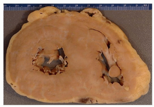
Grossly, the heart at autopsy is generally enlarged (Fig. 148.1) and of firm consistency, and with advanced amyloid shows a waxy beaded endocardial surface of the atria and of the atrioventricular valves, which have propensity for the deposits. The mean heart weight at autopsy is about 550 g.13 In autopsies of cases of AL-type amyloidosis, there is typically gross enlargement of the spleen (Fig. 148.2). The gross consistency of the myocardium is often described as firm, rubbery or waxy. Atrial dilatation is a constant feature (owing to the underlying restrictive hemodynamics), but ventricular dilatation is uncommon. In cases of AL-type cardiac amyloidosis, either primary or secondary to multiple myeloma, gross involvement of viscera, especially liver and spleen, may occur. The hypertrophy resulting from amyloid may occasionally result in septal asymmetry, mimicking hypertrophic cardiomyopathy both by cardiac imaging as well as pathologically14 (Fig. 148.1).


FIGURE 148.1 ▲ Cardiac amyloidosis. A short-axis section demonstrates ventricular hypertrophy with septal asymmetry. The heart was an explant from a patient with cardiomyopathy; a diagnosis of amyloid was made first at pathologic examination of the explanted heart.


Microscopic Findings

The histologic appearance of cardiac amyloid is identical to that of amyloidosis of other organs (Figs. 148.3, 148.4, 148.5, 148.6, 148.7, 148.8, 148.9, 148.10, 148.11). Namely, the proteinaceous deposits show reactivity with Congo red stain and exhibit birefringence when placed in cross-polarized light (Fig. 148.12). In native heart biopsies, amyloid is the most common specific diagnosis rendered, and there should always be a high index of suspicion, especially if there is a clinical suggestion. On low magnification, there is patchy, haphazard deposition of eosinophilic to amphophilic

homogenous material (Fig. 148.4) that may exhibit a cracked appearance. In some examples, there may be only interstitial amyloid that may be focal, necessitating careful review of all biopsies (Fig. 148.5). There may be associated myofiber disarray, interstitial fibrosis, and/or hypertrophy (Fig. 148.6). Occasionally, as in other organs, a foreign body-type giant cell reaction is present (Fig. 148.7). The differential diagnosis is fibrosis, which is generally not as randomly distributed, and shows a more fibrillar appearance.

Only gold members can continue reading. Log In or Register to continue

Stay updated, free articles. Join our Telegram channel

Aug 19, 2016 | Posted by in CARDIOLOGY | Comments Off on Cardiac Amyloidosis

Full access? Get Clinical Tree

Get Clinical Tree app for offline access