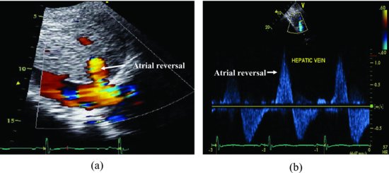
Figure 61.2 The subcostal view with color Doppler shows the blood flow is reversed into the hepatic vein during the systolic period of ventricles (a); the Doppler recording is consistent with the color flow (b). These are typical signs of severe tricuspid regurgitation.
The pulmonic valve was thickened with mild pulmonic regurgitation. (Videoclip 61.2). The estimated gradient of pulmonary artery was 54 mmHg.
Discussion
Stay updated, free articles. Join our Telegram channel

Full access? Get Clinical Tree


