(1)
Ospedale Civile di Vigevano, Vigevano, Italy
Intraventricular Conduction Delays: General Principles
For a review of the anatomy of the His-Purkinje network, see Chap. 1.
Bundle-branch blocks (BBBs) and fascicular blocks (also referred to as hemiblocks) are disturbances involving the electrical activation of the ventricles. As the terms suggest, the conduction impairment usually occurs in the bundle branches or their fascicles, but blocks may also be located in the bundle of His or in the most peripheral branches of the conduction network. Bundle-branch blocks are manifested on the electrocardiogram (ECG) by widening of the QRS complex and may be complete (QRS duration of >120 ms) or incomplete (QRS duration between 110 and 120 ms). The abnormal ventricular depolarization causes secondary changes in the repolarization phase, including inversion of the T waves in leads where they are normally positive and sometimes ST-segment depression.
Bundle-branch blocks can be permanent or temporary, regressing, that is, when the conditions that generated them (e.g., acute ischemia, acute cor pulmonale) begin to improve. They can also be described as stable or intermittent. The latter forms usually appear when the heart rate increases (rate-dependent bundle-branch blocks).
Right Bundle-Branch Block
In the presence of a right bundle-branch block (RBBB), the interventricular septum depolarizes normally, from left to right, and the left ventricle is also activated normally. In contrast, depolarization of the right ventricle is delayed since the right ventricle is the last to be activated. The widening of the QRS complex is largely due to the delayed activation of the right side of the septum and free wall of the right ventricle.
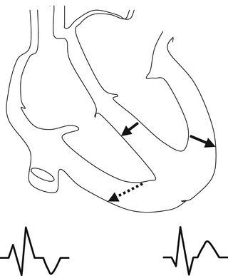
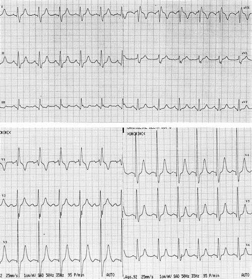
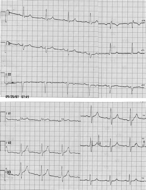
The typical ECG features of RBBB are recorded in lead V1. The normal activation of the septum is inscribed as a large R wave followed by an S wave, which reflects activation of the left ventricle, and a terminal R′ wave representing delayed right ventricular depolarization proceeding anteriorly and to the right (Fig. 8.1). The depth of the S wave in lead V1 varies depending on whether the left ventricular activation vector is oriented posteriorly or anteriorly. In the former case, the R and R′ waves will be separated by a prominent S wave; in the latter case, the S wave may be small, slurred, or completely absent (Figs. 8.2 and 8.3). The leads facing the left side of the septum (I, aVL, V5, and V6) will record an initial q wave followed by an R wave of normal duration and an S wave that is wide and relatively shallow. A late R wave will be present in lead aVR, reflecting delayed activation of the right ventricle.


Fig. 8.1
Diagram showing the altered sequence of ventricular activation during complete RBBB. The solid arrows indicate preserved activation of the interventricular septum and the free wall of the left ventricle. The dotted arrow highlights the delayed activation of the right side of the interventricular septum and free wall of the right ventricle

Fig. 8.2
ECG recording from a patient with severe arterial hypertension and a complete RBBB. In lead V1 there is an rsR′ complex and in leads V6 and I a delayed prominent S wave. The electrical axis is semivertical. The T wave is positive in lead I and negative in V1. Small q waves are present in the leads exploring the left ventricle. The tracing meets the voltage criteria for left ventricular hypertrophy associated with left atrial enlargement

Fig. 8.3
RBBB associated with LAFB. An RR’ complex is present in lead V1
In the presence of a RBBB, the mean axis of the QRS complex in the frontal plane is generally normal. In some cases, there is a right deviation of 15°–30°, but the axis is often indeterminate.
The altered ventricular depolarization process leads to changes in the repolarization phase as well. The direction of the T wave will be opposite to that of the terminal component of the QRS complex, that is, positive in lead I, where the ventricular complex ends with an S wave, and negative in V1, where the terminal wave is an R or R′. If this relation is not maintained, the repolarization abnormality is probably primary rather than secondary.
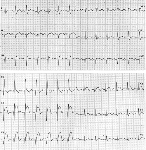
Since the most diagnostically significant elements of the QRS complex are unaltered in RBBB, normality and abnormality can be defined by criteria related to voltage, R-wave progression, and Q waves: RBBB does not interfere with the diagnosis of left ventricular hypertrophy (LVH) or myocardial infarction (Figs. 8.2–8.4).


Fig. 8.4


ECG showing acute myocardial infarction of the anterior wall with complete RBBB and left axis deviation, which can be easily diagnosed on the basis of the QR complexes in V1, the RS complexes in leads I and aVL, and the Rs complexes in lead V6
Stay updated, free articles. Join our Telegram channel

Full access? Get Clinical Tree


