Bulging of the Cardiac Silhouette
A 70-year-old man who had undergone a cardiac transplant 4 months previously suffered a loss of vision in the left eye, but he had no cardiac symptoms or abnormal findings upon physical examination. His chest x-ray showed a bulging of the cardiac silhouette to the right. An echocardiogram was ordered to further evaluate his cardiac status (Fig. 59-1 and Video 59-1).
 Figure 59-1. |
 Figure 59-2. |
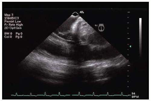 Figure 59-3. |
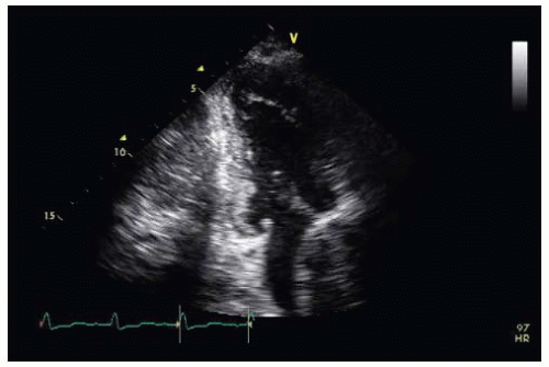 Figure 59-4. |
 Figure 59-5. |
QUESTION 1. This parasternal long-axis image (Fig. 59-1 and Video 59-1) shows:
A. A mass in the left atrium
B. A clot in the left atrium
C. Compression of the left atrium
 Figure 59-6. |
 Figure 59-7. |
 Figure 59-8. |
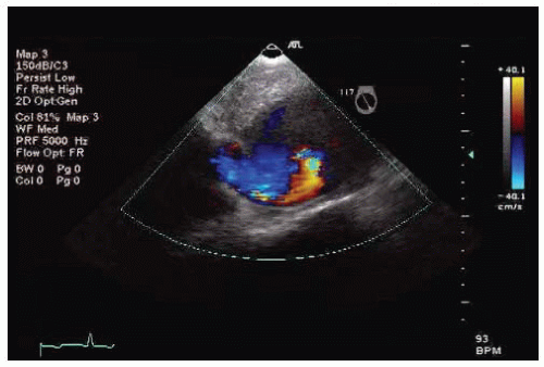 Figure 59-9. |
QUESTION 2. What imaging test do you order next?
A. Transesophageal echocardiogram (TEE)
B. Magnetic resonance imaging (MRI)
C. Computed tomography (CT)
D. Any of the options
View Answer


ANSWER 2: D. As the compression of the atrium is superior, it needs an imaging modality that allows good assessment of the left atrium as well as the aorta. TEE, CT, and MRI are all sufficient for this purpose.
Stay updated, free articles. Join our Telegram channel
Full access? Get Clinical Tree
