Bronchial Masses in the Pediatric Population
Megan K. Dishop MD
Endobronchial biopsy is occasionally performed for diagnosis and/or excision of obstructive tracheal and endobronchial masses. Most bronchial lesions in children are benign, including predominantly nonneoplastic lesions, such as granulation tissue (sometimes related to foreign body aspiration), pyogenic granulomas, granulomatous infection, hamartomas, and juvenile-onset recurrent respiratory papillomatosis. Recurrent respiratory papillomatosis is typically caused by infection with human papillomavirus types 6 and 11 acquired during delivery, and it results in multiple recurrent squamous papillomas of the larynx, tracheobronchial tree, and, less commonly, the esophagus and pulmonary parenchyma. Frequent excisions are required for recurrent papillomas due to obstructive symptoms and rare occurrence of malignant progression.
Other endobronchial tumors include hemangiomas, myofibroblastic tumors, histiocytic lesions, bronchial neuroendocrine tumors (carcinoid tumor), adenoid cystic carcinoma, mucoepidermoid carcinoma, benign mucous gland adenoma, leiomyoma, and rarely metastatic tumors with an endobronchial component. Bronchogenic carcinoma has been reported in children but is exceptionally rare. Inflammatory myofibroblastic tumors account for approximately 20% of all primary lung tumors in children, most commonly forming intraparenchymal masses and only occasionally presenting as an endobronchial mass. Bronchial carcinoid tumors are the most common bronchial neoplasm in childhood, typically presenting in adolescents as endophytic polypoid masses with variable mural and extrabronchial extension. Although rare, carcinoid tumors account for approximately 80% of primary malignant lung tumors in children. They are considered low-grade malignancies with potential for local recurrence and distant metastasis.
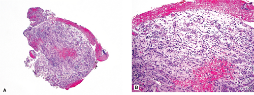 Figure 39.1: Endobronchial biopsy: granulation tissue. A 10-year-old boy presented with recurrent pneumonia and possible bronchial foreign body. Excisional biopsy of an endobronchial lesion (A) shows vascular proliferation in a myxoid inflammatory background, associated with surface hemorrhage (B), typical of granulation tissue. |
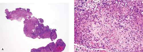 Figure 39.2: Endobronchial biopsy: necrotizing granulomatous inflammation. A 15-month-old boy presented with wheezing and a left bronchial lesion. Endobronchial biopsy (A) demonstrated chronic inflammation and epithelioid granulomas with focal central necrosis (B). Although no organisms were detected on special stains or culture, he was treated presumptively for mycobacterial infection.
Stay updated, free articles. Join our Telegram channel
Full access? Get Clinical Tree
 Get Clinical Tree app for offline access

|