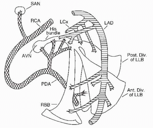
III. SINUS NODE DYSFUNCTION.
Sinus node dysfunction encompasses any dysfunction of the sinus node and includes inappropriate sinus bradycardia, SA exit block, SA arrest, and tachycardia-bradycardia syndrome.
A. Clinical presentation.
There is a wide range of presentations, and some patients’ disease may be asymptomatic.
1. Syncope and presyncope are the most dramatic presenting symptoms. Fatigue, angina, and shortness of breath are more subtle consequences of sinus node dysfunction.
2. In the tachycardia-bradycardia syndrome, the primary complaint may be palpitation. Documentation of the arrhythmia may be difficult because of the sporadic and fleeting nature of the problem.
C. Electrocardiographic findings
1. Inappropriate sinus bradycardia, also known as “chronotropic incompetence,” is defined as a sinus rate of < 60 beats/min that does not increase appropriately
with exercise. Inappropriate sinus bradycardia must be differentiated from a low resting heart rate, which may be normal in athletes and sleeping individuals.
2. Sinus arrest, or sinus pause, occurs when the sinus node fails to depolarize on time. Pauses of < 3 seconds may be seen on Holter monitoring in up to 11% of normal adults (especially athletes) and are not a cause for concern. However, pauses lasting longer than 3 seconds are generally considered abnormal and are suggestive of underlying pathology, especially if the patient is awake when they occur.
3. SA exit block, although similar to sinus arrest on the electrocardiographic tracing, may be distinguished by the fact that the duration of the pause is a multiple of the sinus PP interval. High-grade SA exit block cannot be differentiated from prolonged sinus arrest and is treated in the same manner.
4. Tachycardia—bradycardia syndrome, also referred to as “sick sinus syndrome,” is characterized by episodes of sinus or junctional bradycardia interspersed with an atrial tachycardia, usually paroxysmal atrial fibrillation.
D. Diagnostic testing.
Invasive testing is used when noninvasive methods have failed to yield a diagnosis and sinus node dysfunction is still strongly suspected.
1. Noninvasive testing
a. Electrocardiogram (ECG). In evaluating sinus node dysfunction, the initial workup should include a 12-lead ECG, followed by a 24-hour to 48-hour ambulatory ECG monitoring, if necessary. Use of a diary during the record ing period can help correlate symptoms with the cardiac rhythm. For less frequent events, a loop recorder or an event recorder may be used to assess symptoms over a 2-week to 4-week period. Stress testing can help document the severity of chronotropic incompetence.
b. Autonomic testing includes physical maneuvers, such as carotid sinus mas sage and tilt table testing, as well as pharmacologic interventions to test the autonomic reflexes.
(1) Carotid sinus massage distinguishes intrinsic sinus pause/sinus arrest from a pause due to
carotid sinus hypersensitivity, which is a 3-second
or longer pause and/or a ≥ 50 mm Hg or greater drop in blood pressure that occurs with massage of the carotid sinus (firm pressure applied to one carotid sinus at a time for 5 seconds).
Carotid sinus massage should not normally precipitate sinus pause/sinus arrest, although it will decrease the rate of depolarization of the SA node and slow conduction in the AV node.
(2) Tilt table testing may help differentiate between syncope caused by sinus node dysfunction and that due to autonomic dysfunction. Bradycardic episodes precipitated by tilt table testing are usually caused by autonomic dysfunction and not by sinus node dysfunction.
(3) Pharmacologic testing may be used to differentiate between sinus node dysfunction and autonomic dysfunction. Total autonomic blockade is achieved after administration of atropine 0.04 mg/kg and propranolol 0.2 mg/kg. The resulting intrinsic heart rate represents the sinus node rate, devoid of autonomic influences. Assuming that the normal intrinsic heart rate (in beats/min) is defined by the formula
then an intrinsic heart rate lower than predicted using this formula is consistent with sinus node dysfunction; an intrinsic heart rate close to the predicted rate in a patient with a clinical presentation of sinus node dysfunction is suggestive of an autonomic dysfunction as a cause of the bradyarrhythmia.
2. Invasive testing.
The two most common tests use indirect measurements of SA node function. Direct measurement of SA node function is laborious and rarely performed.
a. Sinus node recovery time (SNRT) is the time it takes the SA node to recover following paced overdrive suppression of the node.
(1) A delay of longer than 1,400 milliseconds is considered abnormal. This measurement may be corrected by subtracting the intrinsic sinus cycle length (in milliseconds) from the recovery time. A corrected SNRT > 550 milliseconds is suggestive of sinus node dysfunction.
(2)The limitations of this test are as follows:
(a) It is an indirect measurement of SA node function and reflects both sinoatrial node conduction time(SACT) and automaticity.
(b) It may be falsely shortened by an SA node entrance block during atrial pacing (due to failure of the paced impulse to reset the sinus node) or falsely prolonged by an SA node exit block (the sinus node is normal but the impulse cannot leave the node), which affects its specificity.
(c) The SNRT is not prolonged in all patients with sinus node dysfunction, which affects its sensitivity.
b. Sinoatrial node conduction time
(1) The steady-state atrial rate is determined (A1-A1 interval or the time between P waves). Then premature atrial extra stimuli (A2) are introduced by pacing high in the right atrium, starting in late diastole at progressively shorter intervals until atrial refractoriness is found (i.e., A2 does not result in a P wave). The duration before the next spontaneous atrial impulse (A3) is measured and the baseline rate is subtracted.
(2) The test assumes that SA node automaticity is not affected by pacing, that con duction time into the node is equal to conduction time out of the node, and that there is no shift in the principal pacemaker site.
E. Therapy.
Treatment for symptomatic sinus node dysfunction may be pharmacologic, pacing, or a combination of both.
1. Indications for pacing in sinus node dysfunction are determined by symptoms (e.g., correlation with a documented arrhythmia;
Table 22.3). Another common indication is when drug therapy that causes sinus node dysfunction cannot be stopped or changed.
2. Medications that suppress sinus node automaticity should be stopped if possible. If this is not possible, it may be necessary to place a temporary or permanent pacemaker (
Table 22.3).
3. For patients with tachycardia-bradycardia syndrome, a pacemaker is often placed for management of the bradyarrhythmia, and antiarrhythmic drugs are added for treatment of the tachycardia episodes.
4. Acute treatment for patients with symptomatic sinus node dysfunction includes the following:
(a) Atropine (0.04 mg/kg intravenous bolus)
(b) Temporary pacing for patients whose conditions fail to respond to drug therapy
(c) Isoproterenol (starting at 1 µg/min intravenously), which may be used as a bridge to pacemaker placement. Isoproterenol is not indicated in most patients with cardiac arrest


