Blood Vessels: Anatomy and Physiology
I. ANATOMY OF BLOOD VESSELS
A. Three Layers (Tunicae) of Blood Vessels
(Fig. 1.1)
1. Tunica interna or intima. This is the innermost layer and is composed of endothelial cells.
2. Tunica media. This is the middle layer and is composed of smooth muscle and elastic fibers. It is thicker in arteries, can change the size and shape of arteries, gives arteries their rigidity and round shape, and is influenced by hormones and other chemicals.
3. Tunica externa or adventitia. This is the outer layer and is composed of collagenous and elastic fibers. It protects and anchors the vessel to surrounding tissues.
B. Circulatory System
(Fig. 1.2)
1. Systemic circulation refers to the flow of blood from the left ventricle of the heart through the body (except for the lungs) and back to the right atrium of the heart. The blood carries oxygen and nutrients to the tissues of the body. It also removes wastes, carbon dioxide, and heat from the tissues of the body. The blood leaves the left ventricle of the heart, and goes through the aorta, arteries, arterioles, venules, veins, and vena cava to enter the right atrium of the heart:
Heart → Aorta → Arteries → Arterioles → Capillaries → Venules → Veins → Vena cava → Heart
2. Pulmonary circulation refers to the flow of blood from the right ventricle of the heart, through the right and left pulmonary arteries, to the alveoli (air sacs) in the lungs, then from the alveoli of the lungs, through the right and left pulmonary veins, and back to the left atrium. The blood is deoxygenated when it enters the alveoli from the right ventricle (as it has already gone through the rest of the body through the systemic circulation) and is oxygenated when it leaves the alveoli of the lungs to go into the left atrium.
Heart → Pulmonary arteries → Alveoli → Pulmonary veins → Heart
C. Types of Blood Vessels
1. Arteries are blood vessels that transport blood from the heart to the tissues of the body. They contain all three layers of tunicae. The tunica interna and media is thicker than in veins. The tunica externa is thinner than in veins. These divide into smaller and smaller branches, eventually dividing into arterioles.
2. Arterioles. These small vessels are regulators of blood flow from the arteries into the capillaries. As they get closer to the capillaries, the layers of arteriole decrease to consist only of an endothelial layer surrounded by a few smooth muscle fibers. Vasoconstriction (when the smooth muscle constricts) decreases blood flow into the capillaries. Vasodilation (when the smooth muscle relaxes) increases blood flow into the capillaries. Arterioles have the highest resistance in the circulatory system. They account for one half of the total resistance to blood flow.
3. Capillaries. These microscopic vessels only have a single layer of endothelium and a basement membrane. They allow exchange of nutrients and waste products between the blood and the cells of tissue. Capillaries (sometimes extensive networks of capillaries) usually connect arterioles and venules.
4. Venules. These vessels drain blood from the capillaries into the veins. Close to the capillaries, venules may only consist of an endothelial layer surrounded by the tunica externa. Closer to the veins, venules consist of all three layers.
5. Veins. Veins are blood vessels that transport blood from the tissues of the body back to the heart. They are composed of all three layers of tunicae, although the tunica intima and tunica media are thinner than in arteries. The tunica externa is thicker than in arteries. Veins contain valves to prevent backflow of the blood, which has lower pressure at this point.
6. Vasa vasorum. This is a network of minute blood vessels that perfuse the tissues of blood vessels themselves.
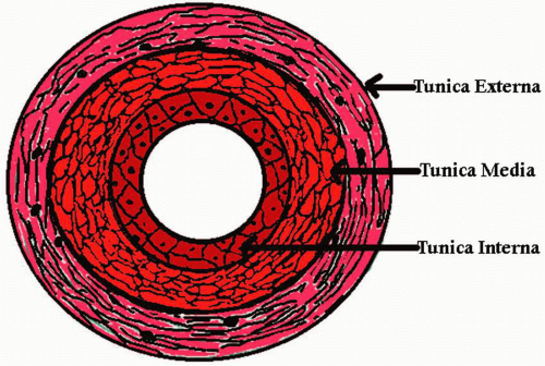 FIGURE 1.1. Three layers of a vessel wall. |
II. PHYSIOLOGY AND CHARACTERISTICS OF BLOOD FLOW
A. Blood flow
is the amount of blood that passes through a vessel during an episode of time. Blood flows in a laminar flow pattern in most vessels (Fig. 1.3). A laminar flow pattern is a stable pattern consisting of many laminae (layers) that are concentric. Each layer is thought to flow at a different velocity. The velocity of each layer increases as they approach the center of the lumen. The center is where the highest velocity of blood flow is thought to exist. The two primary factors that determine blood flow are blood pressure and resistance.
Stay updated, free articles. Join our Telegram channel

Full access? Get Clinical Tree


