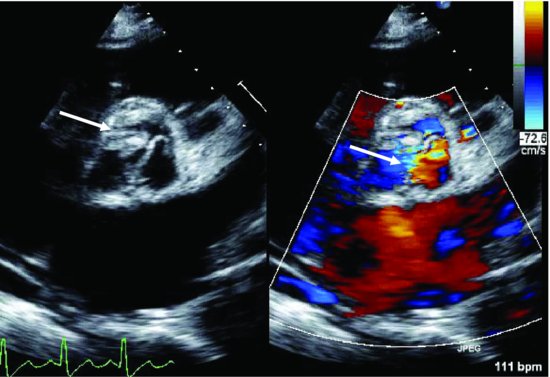
Figure 14.2 The vegetations on aortic valve are seen in the PSA view (arrow); the AR is demonstrated by color Doppler (arrow).

The echocardiography was performed at the time of this admission: The aortic valve bioprosthesis was normal (Figure 14.3 and Videoclip 14.2). Severe periprosthetic posterior directed jet of MR (Figure 14.4 and Videoclip 14.3). This jet is related to mitral prosthetic valve dehiscence. No vegetations were seen. The LV cavity was markedly increased in size with reduced contractility. Transesophageal echocardiography (TEE) revealed: no evidence of endocarditis. Dehiscense of the mitral valve was seen on both 2D and 3D images (Figure 14.5 and Videoclip 14.4). The aortic bioprosthesis valve is functioning well; there was no evidence of aortic valve endocarditis.


