Benign Lung Tumors

Chapter 68 Benign Lung Tumors*


When used together, the words “tumor” and “lung” suggest “malignancy,” as expected given the frequency and lethality of lung cancer worldwide. The work of the respiratory medicine physician and the surgeon would be simpler if all focal opacities in the lung were malignant, but in reality, more are benign than malignant, requiring that the two be separated. A tumor is defined as abnormal benign or malignant growth, possessing no physiologic function, and arising from uncontrolled cellular proliferation. In a strict sense, “inflammation” is both physiologic and usually controlled, and thus does not fit this definition. However, in everyday practice, physicians know a focal opacity is present on imaging without knowing whether it is “physiologic” or “uncontrolled.” This chapter discusses the general approach to benign lung tumors and the most common benign neoplastic and non-neoplastic causes.


Detection and Diagnosis


We tend to use the terms tumor and nodule (or mass) synonymously. Benign tumors may also present primarily in the airway versus the lung parenchyma, and certain causes may present in either location or in both locations. Patients presenting with symptoms and found to have tumors on chest x-ray films or computed tomography (CT) imaging studies are more likely to have a lung malignancy.


Incidentally found tumors or nodules in the lung detected at CT screening are more likely to be benign. Studies of CT screening for lung cancer detect one or more nodules in about 25% of participants when CT collimation (slice thickness) is 10 mm and 40% to 60% of participants at 5 mm or less; about 98% of these nodules are benign. Nodule size is generally an excellent guide for determining benign from malignant; less than 1% of nodules 5 mm or less in diameter represent malignancy, even in current or former smokers. This distinction becomes more difficult for larger nodules, with the likelihood of malignancy more than 50% for nodules 2 cm in diameter, and increasing with larger nodule/mass size. A few benign tumors, such as hamartomas and teratomas, may have imaging features such that CT is diagnostic. However, most benign tumors do not have signature characteristics on imaging, and histology is required for diagnosis.


Positron emission tomography (PET) can be helpful in identifying benign from malignant tumors because it is based on the principle that cancer cells have a high rate of glycolysis compared with non-neoplastic cells. False-positive PET scans have been reported with infections, sarcoidosis, and other benign conditions. False-negative PET scans may occur with low-grade tumors, carcinoid tumor, and malignancies less than 1 cm in diameter. PET is now most often performed with integrated CT (PET-CT). Avidity on PET-CT is not the same as tissue, and a biopsy should be obtained rather than assuming a diagnosis or a stage.


Once a nodule is 3 cm or larger, and it is not clearly benign by showing evidence of calcification or fat on CT, the likelihood of malignancy is greater than 90%. Therefore, most benign tumors larger than 3 cm are diagnosed at resection because of the high index of suspicion of malignancy. Although most inflammatory lesions show no evidence of growth in follow-up, many benign tumors will grow and prompt concern for malignancy and subsequent removal for diagnosis. Bronchoscopy or transthoracic needle biopsy may be used to diagnose benign tumors, especially when multifocal or complete resection is not feasible.


Obstruction of the trachea and major bronchi is most often caused by squamous cell and small cell carcinoma or carcinoid tumor, but tracheal obstruction may also be caused by benign tumors. Several benign tumors manifest more frequently in the airways rather than the periphery of the lung. The patients may have symptoms of airway obstruction, such as cough, recurrent infection, wheezing, and dyspnea. The endoscopic appearance of a lesion may suggest a diagnosis, but uniformly, histology is required. If the lesion is polypoid and has a low likelihood of malignant behavior, successful treatment may be achieved with endoscopic techniques. Broad-based lesions and those with greater malignant potential are best treated with surgical excision; lung-sparing procedures such as a bronchoplasty or sleeve resection may be appropriate.


Benign Epithelial Neoplasms


Papillomas


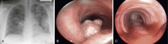
Benign epithelial neoplasms are generally rare, although squamous papilloma is the most common. Histologically, these are identified as papillary tumors with a squamous cell epithelial surface and delicate connective tissue attachments. They may be solitary or multiple and most often occur in the larynx and trachea, with less than 10% having lower airway involvement and only 2% within the lung parenchyma (Figure 68-1). The squamous type of papilloma has an association with human papillomavirus (HPV types 16, 18, 31, 33, and 35). Obstructive symptoms may develop from airway involvement and are an indication for laryngoscopic or bronchoscopic removal. Recurrent papillomas occur in as many as 20%, and some patients require periodic endoscopic debridement. Malignant transformation to squamous cell carcinoma may occur. Compared with squamous cell papillomas, glandular and mixed-cell papillomas are exceedingly rare.


Jun 12, 2016 | Posted by in RESPIRATORY | Comments Off on Benign Lung Tumors

Full access? Get Clinical Tree

Get Clinical Tree app for offline access