Bell-Shaped Chest
Christopher M. Walker, MD
Stephen L. Done, MD
DIFFERENTIAL DIAGNOSIS
Common
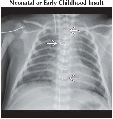
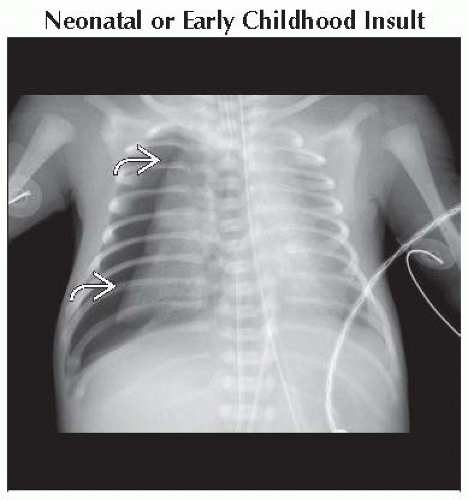
Neonatal or Early Childhood Insult
Less Common
Down Syndrome
Muscular Disorders
Rare but Important
Rickets
Intrauterine Oligohydramnios
Skeletal Dysplasia
Giant Omphalocele
ESSENTIAL INFORMATION
Key Differential Diagnosis Issues
Definition
Narrow upper chest with outward bowing of lower chest
Obliquely directed posterior ribs
Caused by disorders resulting in cerebral or muscular dysfunction
Clinical history key in diagnosis
Helpful Clues for Common Diagnoses
Neonatal or Early Childhood Insult
Conditions leading to hypotonia
Sepsis, dehydration, infantile respiratory distress syndrome, spinal cord injury, and intracranial hemorrhage
Transient occurrence secondary to traversing birth canal
Paralytics for mechanical ventilation
Helpful Clues for Less Common Diagnoses
Down Syndrome
Hypotonia and bell-shaped thorax ↓ in severity with age
Thoracic skeletal manifestations
Bell-shaped chest in 80%
Multiple manubrial ossification centers in 80%
11 rib pairs in 30%
58% chance of Down syndrome with above 3 findings
Gastrointestinal malformations
Duodenal atresia/web, tracheoesophageal fistula, omphalocele, Hirschsprung disease, and imperforate anus
Cardiac malformations
Endocardial cushion defect, ventricular septal defect, atrial septal defect, tetralogy of Fallot, and patent ductus arteriosus
Atlantoaxial instability in 14% of patients secondary to laxity of transverse ligaments
Diagnose with flexion/extension radiography
Muscular Disorders
Spinal muscular atrophy
Progressive lower motor neuron degeneration
Multiple subtypes with varying ages of presentation
Autosomal recessive
Recurrent respiratory infections
Chest shape progressively ↑ in severity with most extreme example of bell-shaped thorax
Muscular dystrophy
Primary muscle breakdown
Varying age of presentation and disease severity depending on type of muscular dystrophy
± rapidly progressive scoliosis
C-shaped scoliosis leads to respiratory insufficiency
Treat with long-segment fusion
Helpful Clues for Rare Diagnoses
Rickets
Widened growth plates
Osteomalacia
Metaphyseal cupping and fraying
Deformed costochondral junctions (rachitic rosary)
Intrauterine Oligohydramnios
Long-term oligohydramnios leads to pulmonary hypoplasia with muscular hypotonia
Causes
Renal abnormalities, urinary bladder obstruction, posterior urethral valves, or asymmetric growth retardation
Fetal ultrasound
Amniotic fluid index (AFI) is defined by 4 largest AP fluid pockets in each quadrant
AFI ≤ 6 cm is diagnostic of oligohydramnios
Skeletal Dysplasia
Abnormal skeletal development with muscular hypotonia
Diagnosis important for prognosis and genetic counseling
Skeletal survey useful in diagnosing and classifying skeletal dysplasias
Important definitions
Rhizomelia implies proximal limb shortening (humerus and femur)
Mesomelia implies middle limb shortening (radius/ulna and tibia/fibula)
Acromelia implies distal limb shortening (hands and feet)
Achondroplasia
Most common short-limbed dwarfism and nonlethal skeletal dysplasia
Autosomal dominant
Prenatal ultrasound demonstrates rhizomelic shortening
↓ AP vertebral body diameter
↓ interpediculate distance in lower lumbar spine
Tombstone-shaped iliac bones
Frontal bossing
Small foramen magnum
Camptomelic dysplasia
Enlarged skull
± 11 rib pairs
Late ossification of mid-thoracic pedicles
Hypoplastic scapulae
Bowed long bones
Tall and narrow iliac wings
Jeune syndrome
Acromelic shortening
Short ribs with high clavicular position
Cone-shaped epiphyses of middle and distal phalanges
“Trident” acetabulum with 3 downward projecting spurs
Normal spine
± ossified capital femoral epiphyses in infancy
Cleidocranial dysplasia
Abnormal membrane bone development
Autosomal dominant
Absent or dysplastic clavicles
Widened pubic symphysis
Squared iliac wings
Wormian bones
Persistent metopic suture
Thanatophoric dysplasia
In utero or early neonatal death
Sporadic disorder
Rhizomelic shortening
± prenatal ultrasound demonstrates cloverleaf-shaped skull
Polyhydramnios
“Trident” acetabulum
Platyspondyly (a.k.a. flat vertebral bodies)
Short ribs
Telephone-receiver-shaped femurs
Metaphyseal flaring
Giant Omphalocele
Herniated bowel and liver covered by peritoneal lining
High association with cardiovascular abnormalities
Image Gallery
 Frontal radiograph shows median sternotomy wires
 in this 14-day-old patient status post coarctation repair. Bell-shaped thorax is presumed secondary to paralytics from intubation in this 14-day-old patient status post coarctation repair. Bell-shaped thorax is presumed secondary to paralytics from intubation  . .Stay updated, free articles. Join our Telegram channel
Full access? Get Clinical Tree
 Get Clinical Tree app for offline access

|

