Basic Principles of 2D Ultrasound


Basic Principles of 2D Ultrasound


Vincent L. Sorrell


1. Which of the following statements is most accurate?


A. Ultrasound consists of waves of compression and decompression, traveling at a variable speed through a specific medium (such as fat or blood).

B. Ultrasound is considered too high for human hearing (generally >2,000 Hz), and infrasound is too low for human hearing (generally <200 Hz).

C. Ultrasound travels in a “longitudinal” (not sinusoidal) wave oscillating back and forth within the direction the sound was transmitted.

D. Most 2D ultrasound probes used in cardiac imaging consist of a range of 2.5 to 5.0 Hz.

E. Unlike audible frequencies, which are difficult to “hear” around objects, ultrasound is more likely to travel around tissue planes, become distorted, and create artifacts.

View Answer

Answer 1: C. Option A is incorrect since sounds will travel at a fixed speed through a specific medium and not a variable speed. In the body, speed will vary as it interacts with different media.

Option B is incorrect since human hearing ranges from 20 to 20,000 Hz, so “ultrasound” is higher than 20,000 Hz (infrasound is <20 Hz).

Option D is incorrect since most cardiac imaging probes are between 1 and 12 MHz (or 1,000,000 and 12,000,000 Hz).

Option E is incorrect since ultrasound, unlike audible sounds, is less likely to diffract and more likely to travel in a straight line.

Unlike audible frequency sound, ultrasound has several unique characteristics that contribute to its diagnostic use. Ultrasound can be directed as a beam and focused. Ultrasound adheres to the rules of reflection (straight target) and refraction (angled target). Small targets will reflect ultrasound and can be characterized. Unfortunately, when ultrasound hits a gaseous medium, attenuation is severe. This is worse with higher frequencies. During propagation of the ultrasound wave, the particles within the medium vibrate parallel to this line of propagation and create longitudinal waves. An ultrasound propagation wave is therefore best characterized by an area of densely packed particles (compression) alternating with areas of loosely packed particles (rarefaction). This ultrasound wave is commonly demonstrated as a sine wave with peaks (compression) and valleys (rarefaction) for convenience. Within the medium, these minor particle oscillations correspond to subtle changes in pressure, but no genuine particle motion transpires.

The degree of reflection, refraction, and attenuation depend on the acoustic properties of the media. The lungs reflect most of the ultrasound energy resulting in poor penetration. However, when the material property of the lungs changes (e.g., pulmonary edema), the ultrasound characteristics predictably change and provide an opportunity for diagnostic imaging. Soft tissues (and blood) reflect much less of the ultrasound energy allowing increased penetration and greater diagnostic value.


2. Which of the following is considered a disadvantage of ultrasound?


A. Ultrasound is poorly transmitted through a gaseous medium, and attenuation is marked.

B. As ultrasound passes through a medium, particles within this path vibrate parallel to the line of propagation.

C. Sound waves can be characterized by regions of closely related particles within the medium (compression) and regions of loosely related particles (rarefaction).

D. Reflection, refraction, and attenuation are each dependent on the acoustic properties of the medium.

E. As ultrasound moves through the medium, particle oscillations may become marked, and this particle motion may result in tissue heating and injury.

View Answer

Answer 2: A. Option B is an advantage of ultrasound. Since particles vibrate parallel to the line of propagation, longitudinal waves are created allowing for the creation of an adequate 2D image.

Option C is also an advantage of ultrasound since this acoustic mismatch within the medium is what is responsible for creating a diagnostic ultrasonic image.

Option D is important and an advantage of ultrasound since this variable property within the media is predictable and consistent.

Option E is incorrect. Importantly, the particle oscillation is very tiny, and no actual particle motion occurs. It would be extremely rare for tissue heating to occur with routine medical ultrasound.

The degree of reflection, refraction, and attenuation will vary on the acoustic properties of the tissue it is traveling through. If there is much air, such as the lung and bone (with its interfaces), then most of the sound is reflected, and attenuation occurs rapidly. Very dense media reflect most of the ultrasound energy and less dense (soft tissues and blood) allow much more ultrasound energy to propagate and create a diagnostic image.


3. Which of the following is the correct order of the velocity of sound within the medium, from fastest to slowest tissue?


A. Blood, bone, and air

B. Air, bone, and blood

C. Air, blood, and bone

D. Bone, blood, and air

E. Bone, air, and blood

View Answer

Answer 3: D. Velocity is fastest in bone (>4,000 m/s) and slowest in air (<350 m/s). Most soft tissue organs and blood are the same, and blood travels at 1,540 m/s (or 1.54 km/s).

The velocity of sound varies depending on the density of tissue as it travels through the medium. TABLE 2.1 represents the velocity of sound and various tissues.


Table 2.1 Velocity of Sound and Various Tissues


TISSUE


TYPE VELOCITY (M/S)


Air


330


Fat


1,450


Soft tissues


1,540


Blood


1,570


Muscle


1,580


Bone


4,080


4. Match the following terms with the most accurate definition:


A. Gain


1. The transfer of ultrasound energy to the tissue during propagation


B. Intensity


2. The net loss of ultrasound energy during propagation


C. Acoustic impedance


3. The degree of amplification of the returning ultrasound signal


D. Absorption


4. Product of tissue density and the velocity of sound


E. Attenuation


5. Distribution of power within an area, analogous to loudness


View Answer

Answer 4:

A—3

B—5

C—4

D—1

E—2

See TABLE 2.2 of basic definitions related to ultrasound.


5. Which of the following statements regarding the interaction between ultrasound and tissue is the least accurate?


A. The shorter the wavelength, the smaller the structures that can be accurately resolved.

B. The higher the frequency of ultrasound, the higher the spatial resolution.

C. The lower the frequency of ultrasound, the higher the tissue penetration.

D. Attenuation always increases with depth regardless of the frequency or tissue type.

E. The greater the acoustic mismatch, the more the energy is reflected than transmitted.

View Answer

Answer 5: D. Of the choices available, option D is the least accurate statement since it is only partially correct. Although it is correct that attenuation always increases with depth, the degree of attenuation is dependent on the ultrasound frequency and the tissue type. Attenuation can be estimated by the following formula: 0.5 dB/cm/MHz. Given than relationship, it can be assumed that the attenuation for a 33.5-MHz transducer imaging an object at 20 cm depth will be 0.5 × 20 × 3.5 = 35 dB. A 5-MHz transducer will undergo much greater attenuation: 0.5 × 20 × 5 = 50 dB.

The higher the frequency (shorter the wavelength), the smaller the structures that can be accurately resolved. Because a goal of imaging is to characterize tiny structures, high frequencies appear desirable. Unfortunately, the physics of diagnostic imaging is robust with trade-offs. The improvement of one aspect of diagnostic imaging is often wrought with another aspect that must worsen for this improvement. High-frequency ultrasound must sacrifice penetration compared with lower frequencies. Ultrasound energy is lost during propagation through a medium (attenuates) due to absorption, scattering, and reflection. Attenuation always increases with depth. Higher frequencies worsen attenuation. Tissue types also drastically impact the rate of attenuation.


6. Which of the following statements regarding the use of ultrasound gel is most accurate?


A. Acoustic coupling gels must always be used because the transducer footprint might otherwise create friction on the skin with resultant tissue injury from heating.

B. Acoustic coupling gels are required to form an image because otherwise 99% of the ultrasound energy is reflected prior to being transmitted into the skin.

C. The use of gel between the transducer and skin surface lowers the percentage of energy transmitted into the body.

D. Acoustic coupling gel is not mandatory to create an ultrasound image, but helps to enhance the signal-tonoise ratio.

E. The use of gel alters the ultrasound frequency, increasing the penetration of the energy.

View Answer

Answer 6: B. Option A is incorrect since the gel is not used to modify injury. There is a very limited risk of heating, and this is not due to friction between the transducer footprint and the skin.

Option B is the correct answer. Due to the very low acoustic impedance of air, gel is necessary to minimize this intervening air layer.

Option C is incorrect since the use of gel decreases the reflection and increases the penetration of ultrasound energy.


Table 2.2 Basic Definitions Related to Ultrasound


TERM


DEFINITION


Absorption


The transfer of ultrasound energy to the tissue during propagation


Acoustic impedance


The product of the density of the medium and the velocity of sound; differences in acoustic impedance between two media determine the ratio of transmitted versus reflected sound at the interface


Amplitude


The magnitude of the pressure changes along the wave; also, the strength of the wave (in decibels)


Attenuation


The net loss of ultrasound energy as a wave propagates through a medium


Cycle


The combination or sum of one compression and one rarefaction of a propagating wave


Dead time


The time in between pulses that the echograph is not emitting ultrasound


Decibel


A logarithmic measure of the intensity of sound, expressed as a ratio to a reference value (dB)


Duty factor


The fraction of time that the transducer is emitting ultrasound; a unitless number between 0 and 1


Far field


The diverging conical portion of the beam beyond the near field


Frequency


The number of cycles per second, measured in hertz (Hz)


Gain


The degree, or percentage, of amplification of the returning ultrasound signal


Half-layer value


The distance an ultrasound beam penetrates into a medium before its intensity has attenuated to one-half the original value


Intensity


The concentration or distribution of power within an area, often the cross-sectional area of the ultrasound beam, analogous to loudness


Longitudinal wave


A cyclic disturbance in which the energy propagation is parallel to the direction of particle motion


Near field


The proximal cylindrical-shaped portion of the ultrasound beam before divergence begins to occur


Period


The time required to complete one cycle, usually expressed in microseconds (µs)


Piezoelectricity


The phenomenon of changing shape in response to an applied electric current, resulting in vibration and the production of sound waves; the ability to produce an electric impulse in response to a mechanical deformation; thus, the interconversion of electrical and sound energy


Power


The rate of transfer over time of the acoustic energy from the propagating wave to the medium, measured in watts


Pulse


A burst or packet of emitted ultrasound of finite duration, containing a fixed number of cycles traveling together


Pulse length


The physical length or distance that a pulse occupies in space, usually expressed in millimeters (mm)


Pulse repetition frequency


The rate at which pulses are emitted from the transducer, that is, the number of pulses emitted within a period of time, usually 1 s


Resolution


The smallest distance between two points that allows the points to be distinguished as separate


Sensitivity


The ability of the system to image small targets at a given depth


Ultrasound


A mechanical vibration in a physical medium, characterized by a frequency >20,000 Hz


Velocity


The speed at which sound moves through a given medium


Wavelength


The length of a single cycle of the ultrasound wave; a measure of distance, not time


From Armstrong WF, Ryan T. Feigenbaum’s Echocardiography. 7th ed. Philadelphia, PA: Lippincott Williams & Wilkins, 2010.


Option D is incorrect since gel is necessary to create an ultrasound image.

Option E is incorrect. Although ultrasound gel results in increased penetration of energy, this effect is not due to an alteration of the ultrasound frequency.

Acoustic mismatch creates ultrasound echoes that obey the rules of optics for reflection and refraction that depend on the angle of incidence and the magnitude of the acoustic impedance. The use of an acoustic coupling gel is necessary to reduce the air-tissue interface at the skin surface (otherwise, >99% of the ultrasonic energy would be lost). This is due to the low acoustic impedance of air. Gel increases the percentage of energy transmitted into the body.


7. Which of the following tissues is not an example of a specular reflector?


A. Left ventricular endocardium

B. Left ventricular myocardial texture or speckles

C. Pericardium

D. Mitral valve

E. Right ventricular epicardium

View Answer

Answer 7: B. As ultrasound passes through the body, it encounters multiple small and large interfaces that alter the transmission of energy. Specular reflectors are created by targets that are large relative to the ultrasound wavelength. This type of interaction is seen in endocardial and epicardial surfaces, cardiac valves, and the pericardium.

Scattered reflectors are created by targets that are small relative to the transmitted wavelength. Only a very tiny portion of the ultrasound energy is reflected back to the transducer. This type of interaction results in the “speckle” pattern that creates the texture within the myocardium. Without this type of interaction, also referred to as Rayleigh scatterers, the myocardial wall would instead appear as two solid lines (endocardium and epicardium/pericardium) with an echo-free space in between.


8. Based upon the 2D short-axis images shown in FIGURE 2.1, which of the following statements is most accurate?


FIGURE 2.1 From Armstrong WF, Ryan T. Feigenbaum’s Echocardiography. 7th ed. Philadelphia, PA: Lippincott Williams & Wilkins, 2010.


A. The image on the left was obtained using much higher gain than the image on the right.

B. The image on the right has the focus much closer to the near field than the image on the left.

C. The image on the left was obtained with fundamental frequency, and the image on the right was obtained using a harmonic multiband imaging probe.

D. The image on the left used a lower-frequency imaging probe compared with the image on the right.

E. The image on the left has higher spatial resolution than the image on the right.

View Answer

Answer 8: D. This is an example of the impact transducer frequency changes will have on 2D image quality. The parasternal short-axis image on the left was recorded using a 3.0-MHz transducer, while the image on the right, a similar image, was obtained with a 5.0-MHz probe. The higher-frequency image has superior spatial resolution, which is very noticeable within the myocardium where the lower-frequency image appears more “grainy” and the higher-frequency image is more “smooth.” This smooth appearance is due to the ability of the higher spatial resolution to separate “two points very close together” into “two points” as opposed to “one point.” The “points” or targets on this image reside within the heterogeneity of the normal LV myocardium.

Options A-C are incorrect since the focal point, the gain, and the probe were unchanged. Option E is incorrect since the LV myocardial image on the right has a noticeably higher spatial resolution (thus, confirming that Option D is the only correct answer).


9. Which of the following statements regarding 2D image acquisition is most accurate?


A. Bursts, or pulses, of ultrasound are transmitted after a brief excitation of the piezoelectric elements to create a pulsed-wave Doppler image, but continuous transmission is necessary for 2D imaging.

B. Pulse repetition rates between 1 and 2,000 per second are used for M-mode scanning but higher in 2D scanning and usually between 3 and 5,000 per second.

C. Since M-mode scanning has a higher pulse repetition rate than 2D scanning, M-mode has a higher temporal resolution compared to 2D echocardiography.

D. Conventional 2D ultrasound requires very sensitive receivers since up to 50% of the emitted ultrasound energy may be lost (attenuated) prior to reflecting back to the transducer.

E. Pulse repetition frequencies are indeed lower for 2D scanning since the pulses are transmitted across a 90-degree sector scan.

View Answer

Answer 9: B. Option A is incorrect since all ultrasonic imaging requires energy to be transmitted, reflected, and received, and the transducer must act as both a sender and a receiver of bursts or pulses of ultrasound.

Option C is incorrect since 2D scanning must have a higher pulse repetition rate needed to create an image over a large (90-degree) sector. Since M-mode has all of its pulses dedicated to a single raster line, the temporal resolution is far greater, despite having slower pulse repetition rates.

Option D is incorrect since much more than 50% of the emitted energy is attenuated (99%).

Option E is incorrect since 2D requires higher pulse repetition rates to maintain an adequate temporal resolution despite creating such a large sector of data.

To create a 2D ultrasound image, the ultrasound energy must be transmitted, reflected, and received. A short burst of electricity intermittently excites the piezoelectric elements, which creates a pulse of ultrasound that travels into the body while the transducer patiently waits for the signal to return. The pulse repetition rate of M-mode echocardiography is typically 1,000 to 2,000 pulses/s and 3,000 to 5,000 pulses/s to create the 90-degree sector scan associated with 2D. Although the pulse repetition rate is lower for M-mode, all of the pulses are devoted to a single raster line providing a temporal resolution (TR) that is much higher for M-mode compared with 2D echocardiography. To improve the TR of 2D, one can narrow the image sector, and this will greatly improve the TR. Also, decreasing the depth will quicken the time of the transmitted energy to return to the receiver and also improve the TR. Transducers must have very sensitive receivers that can detect signal that are markedly attenuated since <1% of the emitted ultrasound energy is typically reflected.


10. Which of the following relationships regarding the creation of a 2D image is least accurate?


A. The PRF (pulse repetition frequency) is dependent on the depth of the image.

B. Increasing the 2D sector angle from 60 to 90 degrees requires more raster lines to maintain image quality.

C. A line density (# lines/degree) of at least 200 lines/degree is necessary for maintaining high-quality 2D images.

D. Increasing the frame rate will lower the line density and worsen the image quality.

E. To include color-flow images on the same display as 2D images, the entire velocity spectrum cannot be measured, and only the mean frequency (and frequency spread—“variance”) is calculated.

View Answer

Answer 10: C. Answer A is a correct statement since the PRF is greatly dependent on the time to transmit and receive an ultrasound burst. Since the speed of sound is relatively fixed, the most important variable is the depth of the image.

Answer B is a correct statement since more lines of data are necessary to create and maintain an adequate image quality as the sector increases.

Answer C is the least accurate statement since 2 lines/degree are necessary to obtain high-quality images, and it would be prohibitively long to obtain 200 lines/degree.

Answer D is a correct statement. To increase the frame rate, the line density is compromised, and therefore, the image quality degrades.

Answer E is a correct statement. This concept is increasingly important as one tries to compare tissue Doppler velocities obtained from two distinct techniques. Since color coding measures mean velocities, these measures will be inherently lower than if pulsed-wave Doppler measures were obtained.

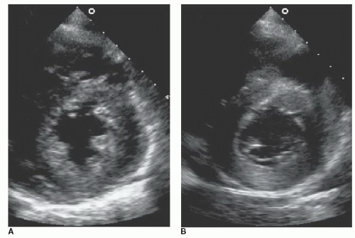
A number of variables must be considered when creating a 2D image. The depth of the examination, the pulse repetition frequency (PRF), the line density, and frame rate each conspire to result in a high-quality image. After sending in a burst of ultrasound, the returning signal must be received, and this is mostly a function of the depth of transmission. Each ultrasound pulse provides a single raster line of data, and the rate that the pulses are transmitted is the PRF. To obtain a 2D image (as opposed to a single-dimensional M-mode image), the ultrasound beam needs to sweep through an angle. The greater the line density, the higher the spatial resolution and image quality. More data lines are needed to fill larger sectors. A line density of approximately two lines per degree is usually adequate to construct a high-quality image. Line density can be increased by decreasing the sector width (same # lines per smaller region), reducing the frame rate (more time for building lines/frame), and reducing the depth (shorter lines = shorter time to build).

The total ultrasonic data recorded during one complete sweep of the ultrasound beam are termed a “field.” The sum of imaging data recorded is a frame. Two fields are interlaced (to improve line density) to produce one frame, and therefore, the frame rate is half of the sweep rate.

Color-flow Doppler (CFD) imaging is a form of pulsed-wave Doppler imaging. CFD uses multiple sample volumes along multiple raster lines to record the Doppler shift at multiple locations. This information is placed on top of a two-dimensional image display, but only the mean frequencies and frequency spreads (variance) are obtained to save processing (Fourier transform) time.


11. You are asked to assist in the performance of an intraoperative TEE as the surgeon is coming off bypass. When you arrive on the scene, the anesthesiologist is frustrated since the US system just “shut down” after an error message about “heating.” Upon further questioning, the same midesophageal image was being viewed by medical students throughout the entire 45-minute operation. Which of the following statements is most accurate?


A. Heating is so rare; this likely represents a malfunction of the ultrasound system or TEE probe. This patient is at potential risk for severe injury, and the probe should not be used again until checked.

B. This likely could have been avoided by maintaining a transgastric view instead of an esophageal view.

C. This likely could have been avoided by using the freeze button or reducing the mechanical index.

D. This is a normal US system process and is likely due to a febrile patient. It requires the operator to “dial up” the “patient temperature” to match the probe temperature.

E. Restarting the US system and continuing to image as before are safe and do not place this patient at an increased risk.

View Answer

Answer 11: C. Option A is incorrect since it is not the single best answer. Although a system malfunction may have occurred, in this situation, a malfunction was not necessary since the students kept the high-frequency TEE probe imaging for 45 minutes at a single location. This invokes a potential risk of esophageal burn. Although not reported in the literature, esophageal perforation has been demonstrated, and the role of localized heating cannot be excluded as a contributing factor.

Option B is incorrect since although the gastric mucosa is thicker (and risk of injury likely less), probe heating will still occur if left imaging in a stationary position for this long.

Option C is the single best answer. Steps to reduce localized heating include limiting the overall imaging time (use freeze button when not actually using US image data) and reduction in imaging power (mechanical index). See discussion below.

Option D is incorrect. Although a febrile patient may actually result in a higher temperature being registered on the TEE probe and US system, assuming this is the cause and increasing the “patient temperature” setting risks potential mucosal injury from heating.

Option E is incorrect since it would pose potential risk to continue to “image as before,” which already resulted in the system’s warning mechanism to operate as designed and shut down once before.

Only gold members can continue reading. Log In or Register to continue

Stay updated, free articles. Join our Telegram channel

Jun 1, 2016 | Posted by in CARDIOLOGY | Comments Off on Basic Principles of 2D Ultrasound

Full access? Get Clinical Tree

Get Clinical Tree app for offline access