Bacterial Pneumonia
Armando E. Fraire MD
Abida Haque MD
Clifford G. Risk MD
Several decades after the introduction of modern antimicrobials, bacterial pneumonia remains a major cause of morbidity and mortality. Two major types of pneumonia are recognized: lobar pneumonia, presenting with a diffuse uniform inflammation of an entire lobe of the lung, and the more frequent bronchopneumonia, in which the inflammation primarily involves the terminal airways and the secondary lobules. Causative organisms include Gram-positive cocci as well as Gram-negative rods. Among the Gram positive, Staphylococcus aureus and Streptococcus pneumoniae predominate. Among the Gram negatives, Klebsiella pneumoniae, Haemophilus influenzae, Pseudomonas organisms, and Escherichia coli are commonly encountered. The histopathological hallmark of pneumonia is an acute inflammatory and/or fibrinous exudate with secondary necrosis of the alveolar epithelium. A biopsy is seldom required for diagnosis, but bronchoscopists may elect to perform a biopsy when attempting to obtain sterile material for culture and sensitivity. In these instances, and depending on whether pneumonia or bronchopneumonia is present, an acute inflammatory process will be evident in biopsy material.
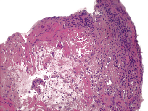
 Figure 10.1: Medium power of transbronchial biopsy shows acute and chronic inflammatory infiltrates in the submucosal tissue in a case of Pseudomonas bronchopneumonia confirmed by microbiologic culture.
Stay updated, free articles. Join our Telegram channel
Full access? Get Clinical Tree
 Get Clinical Tree app for offline access

|