3.2 AV Conduction Disorders
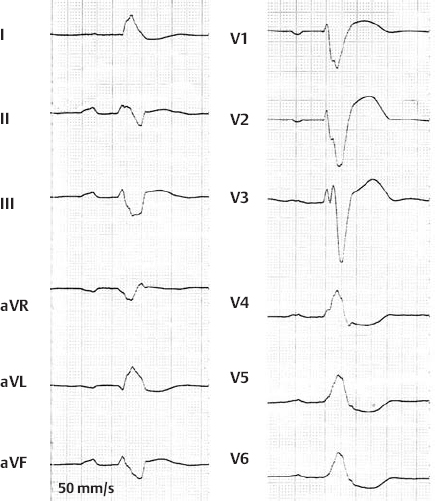
1st Degree AV Block
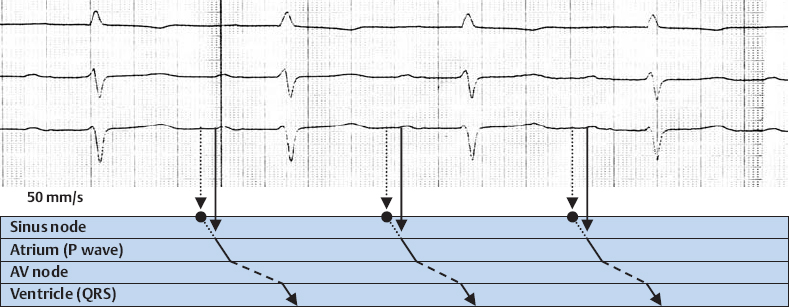
1st Degree AV Block

1st Degree AV Block
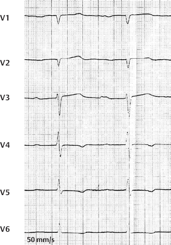
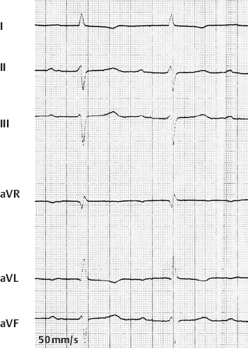
1st Degree AV Block with Complete Left Bundle Branch Block
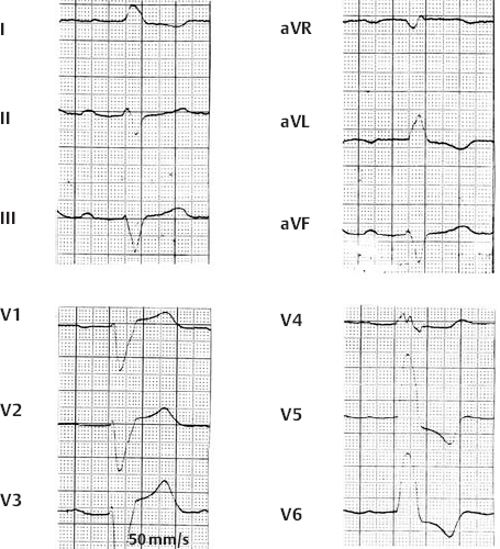
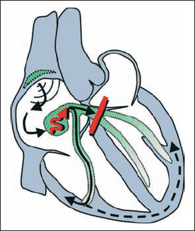
1st Degree AV Block with Complete Left Bundle Branch Block

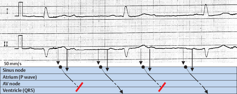
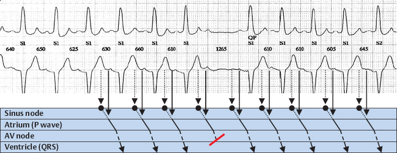
2nd Degree AV Block, Wenckebach Type
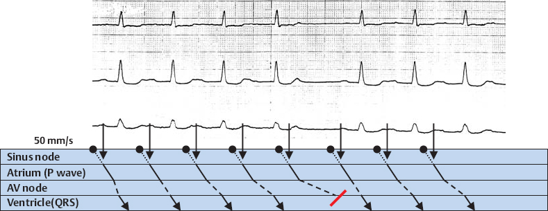
2nd Degree AV Block, Wenckebach Type
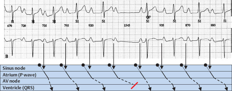
2nd Degree AV Block, Wenckebach Type
2nd Degree AV Block, Wenckebach Type
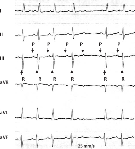

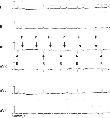
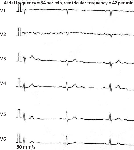
2nd Degree AV Block, 2:1 Block
2nd Degree AV Block, Mobitz Type
2nd Degree AV Block, 2:1 Block

Stay updated, free articles. Join our Telegram channel

Full access? Get Clinical Tree


