Fig. 23.1
(a, b) A third-degree atrioventricular block with slow ventricular escape rhythm (35 bpm) is shown. Atrioventricular dissociation with atrial rate faster than the ventricular rate is clearly visible. QRS has a left bundle branch block (LBBB) pattern
shows a third-degree atrioventricular block with slow ventricular escape rhythm (35 bpm); There is a clear atrioventricular dissociation with atrial rate faster than the ventricular. QRS has a left bundle branch block (LBBB) pattern.
Echocardiogram showed
normal dimensions and function of cardiac chambers (ejection fraction 55–60 %), aortic valve prosthesis normally functioning, mild mitral regurgitation, and mild tricuspid regurgitation with a normal pulmonary artery systolic pressure.
Clinical Course and Therapeutic Management
A continuous electrocardiographic monitoring was placed, and we started isoproterenol infusion that increased HR up to 44 bpm (0.02 mcg/kg/min). After a complete diagnostic workup aimed to exclude secondary reversible causes for AV block, a dual-chamber pacemaker was implanted the day after hospital admission. After implantation ECG showed sinusal P waves followed by paced QRS complexes (Fig. 23.2a, b). The chest X-ray showed the following things: the normal cardiac and pulmonary dimensions, and a correct positioning of the pacemaker leads, and no complications related to the surgery procedure.
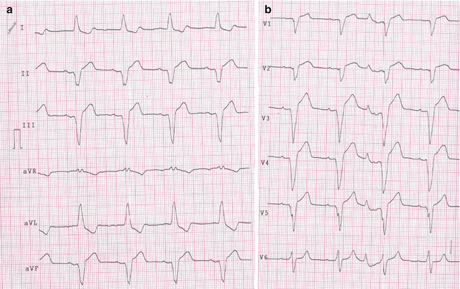
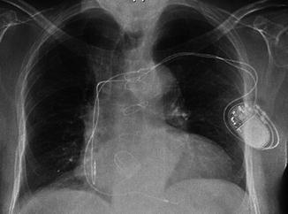
Fig. 23.2
(a) Atrial flutter with third-degree atrioventricular block and junctional escape rhythm. (b) The same patient after cardioversion: sinus rhythm with AV dissociation is shown
Fig. 23.3
Chest X-ray showing normal cardiac and pulmonary dimensions, correct positioning of the pacemaker leads, and no complications related to the surgery procedure
On the 3rd day, the patient was discharged.
A follow-up visit was performed 1 month later. The patient was completely asymptomatic. Physical examination was normal. ECG showed regular rhythm with paced QRS following sinusal P waves (VDD mode). PMK was interrogated and did not show any malfunction.
23.2 Atrioventricular Blocks
An AV block is present when the atrial impulse is conducted with delay or is not conducted at all to the ventricle. The block can occur within the AV node, His bundle, or bundle branches. In the first-degree heart block, conduction time is prolonged but all impulses are conducted. Second-degree heart block is characterized by occasional or repetitive sudden block of conduction of an impulse. In third-degree AV block, no impulses are conducted from atria to ventricles. The term advanced heart block indicates blockage of two or more consecutive impulses. Retrograde conduction can occur in the presence of anterograde AV block [1, 2].
First-Degree AV Block
During first-degree AV block, every atrial impulse conducts to the ventricles and a regular ventricular rate is present. The prolonged PR interval exceeds 200 ms in adults. The conduction delay can be located within the AV node (A–H interval prolonged), in the His–Purkinje system (H–V interval prolonged), or both. If the QRS complex has a normal width, the AV delay most likely resides within the AV node and rarely within the His bundle. On the contrary, if the QRS has a bundle branch block pattern, the conduction delay may be within the AV node or His–Purkinje system as well. Enhancement of vagal tone by carotid massage can cause first-degree AV nodal block to progress to type I second-degree AV block [2].
Second-Degree AV Block
In second-degree AV block, the nonconducted P wave can be intermittent and the preceding PR interval duration may be fixed or prolonged. In this type of block, the P–QRS relationship is not random. Mobitz type I (Luciani–Wenckebach) is characterized by progressive PR prolongation until a nonconducted P wave. In contrast, in Mobitz type II, the PR interval remains constant before a P wave is blocked [3, 4].
During Mobitz type I block, the conduction time delay is longer in the second beat of the Wenckebach group; meanwhile, prolonged conduction time decreases progressively in the subsequent beats. The consequence is that the interval between the successive beats progressively decreases, although the conduction time increases. The RR interval produced by the block is shorter than the double of the RR interval that precedes the block impulse (the shortest interval). The R–R interval that follows the nonconducted beat (the first beat of the new Wenckebach group) is longer than the cycle preceding the blocked impulse [5].
Type II AV block is often a precursor of syncope and complete AV block, because it is more likely to be sub-Hisian. Type I AV block with a normal QRS complex generally does not progress to more advanced forms of AV conduction disturbances. In elderly people, type I AV block may be associated with a clinical course similar to that seen in type II.
In the acute coronary syndromes (ACS), type I AV block generally develops with inferior infarction, is transient, and does not require temporary pacing [4].
Type II AV block occurs usually in the setting of an anterior myocardial infarction, can require temporary or permanent pacing, and is associated with a high mortality rate.
A high-degree AV block can occur also in patients with acute inferior myocardial infarction (MI) and is associated with an extended myocardial damage and a higher mortality rate than those without AV block. All types of AV blocks in the setting of an acute MI can be reversible within 14–21 days.
In type I AV block with a normal QRS duration, the conduction delay is likely to be at the AV node level proximal to the His bundle. A type I intra-Hisian block is uncommon. Type II AV block, particularly in association with a bundle branch block, may be localized within or below the His–Purkinje system. Type I AV block in a patient with a bundle branch block can be caused by a block in the AV node or in the His–Purkinje system as well [2].
Type 2:1 AV block can be due to type I or type II AV block.
Abrupt transient alterations in autonomic tone can also cause sudden block of one or more P waves without altering the PR interval of the conducted P wave before or after the block. Such condition is a typical normal finding at night during sleep. Thus, apparent type II AV block would be produced at the AV node. Increased vagal tone, producing an AV block, usually lengthens the P–P interval at the same time [4].
Vagal stimulation generally increases and vagolytic agents decrease the extent of type I AV block. Atropine can minimally improve conduction in the AV node and markedly faster sinus rate, which may result in higher AV block degree. Counterwise, carotid sinus massage generally improves and atropine worsens AV conduction in patients with a conduction block within or below the His bundle.
Similarly, exercise or isoproterenol by increasing the sinus rate and improving AV conduction may resolve the supra-Hisian block and impair sub-Hisian one [2].
First-degree and type I second-degree AV block can occur in normal healthy children; a Wenckebach AV block can be also normal in well-trained athletes, probably related to a higher vagal tone at rest.
Third-Degree AV Block
In the total AV block, no atrial activity is conducted to the ventricles; the atria and ventricles are controlled by independent pacemakers (atrioventricular dissociation) [4]. The atrial pacemaker can be sinus or ectopic (tachycardia, flutter, or fibrillation) (Fig. 23.4).
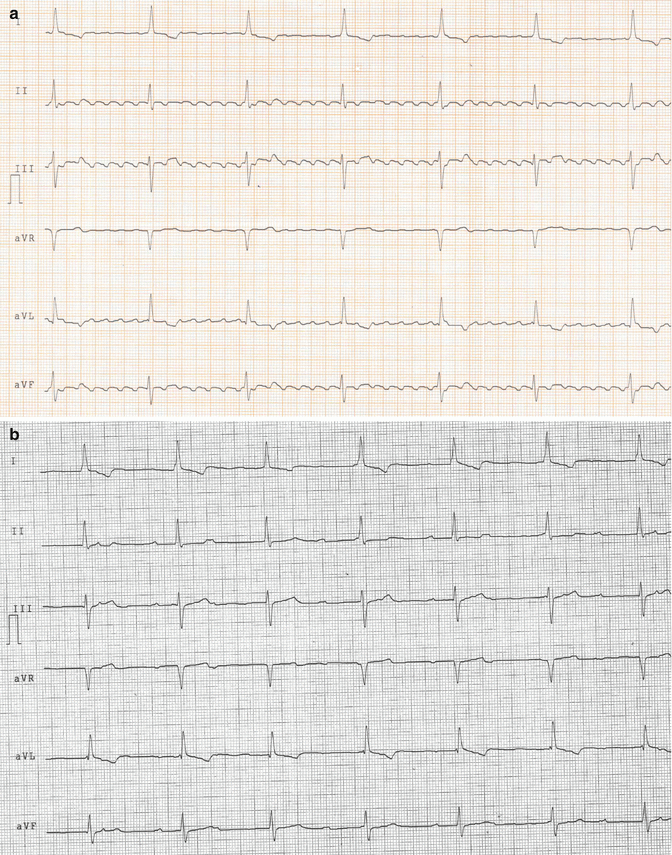
Fig. 23.4
The ventricular rate arise below the region of the block. The atrial pacemaker can be (a) ectopic (tachycardia, flutter, or fibrillation) or (b) sinus
Complete AV block may arise at AV node level (usually congenital), within the bundle of His, or distally below the His (usually acquired).
The ventricular focus that arise below the region of the block. Sites of ventricular pacemaker activity that are in or closer to the His bundle are more stable and can produce a faster escape rate than those located distally in the ventricular conduction system. The ventricular escape rate in acquired total heart block is less than 40 beats/min but can be faster in congenital complete AV block [5].
< div class='tao-gold-member'>
Only gold members can continue reading. Log In or Register to continue
Stay updated, free articles. Join our Telegram channel

Full access? Get Clinical Tree


