Figure 41.2 Transesophageal echocardiogram image shows the contrast through patent foramen ovale into the left atrium and left ventricle. AO, aorta; LA, left atrium; LV, left ventricle.
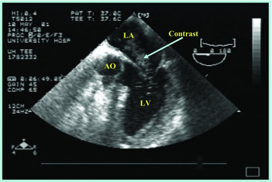
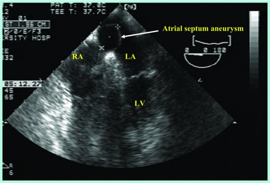
Figure 41.3 The maximal oscillation of atrial aneurysm was 17 mm. LA, left atrium; LV, left ventricle; RA, right atrium.
Discussion
ASA is a localized deformity of the interatrial septum, generally at the level of the fossa ovale, which bulges into the right or left atrium or both. ASA was initially thought to be a rare congenital abnormality; however, with the advent of two-dimensional echocardiography and more recently, with the widespread use of TEE it is more easily and more frequently identified in patients.
The prevalence of ASA varies; TTE studies estimate the rate to be between 0.12 and 0.54% [1]. In a large autopsy series the prevalence reported was 1%. Studies using TEE have demonstrated a prevalence between 3 and 8% [2, 3].
Stay updated, free articles. Join our Telegram channel

Full access? Get Clinical Tree


