Fig. 5.1
Schematic diagram of SCCT coronary segmentation. RCA right coronary artery, R-PDA right posterior descending artery, L-PDA left posterior descending artery, L-PLB left posterolateral branch, R-PLB right posterolateral branch, LAD left anterior descending artery. 1 pRCA, 2 mRCA, 3 dRCA, 4 R-PDA, 5 LM, 6 pLAD, 7 mLAD, 8 dLAD, 9 D1, 10 D2, 11 pLCx, 12 OM1, 13 dLCx, 14 OM2, 15 L-PDA, 16 R-PLB, 17 Ramus, 18 L-PLB
The lumen of the coronary arteries should be examined for overall caliber and smoothness.
Variations in CT density within the mural and intraluminal portions of the coronary artery should be noted and compared with the adjacent interstitium, contrast-containing lumen, and calcific densities such as the bone or calcified plaque.
Atherosclerotic lesions should be considered in relationship to their segmental position due to the affected extent of the myocardium.
The worst lesion should be reported if there are more than two lesions in one segment.
Table 5.1
Explanation of abbreviation of SCCT coronary segmentation diagram
Proximal RCA | pRCA | Ostium of the RCA (right coronary artery) to one-half the distance to the acute margin of heart |
Mid RCA | mRCA | End of proximal RCA to the acute margin of heart |
Distal RCA | dRCA | End of mid RCA to origin of the PDA (posterior descending artery) |
PDA-RCA | R-PDA | PDA from RCA |
PLB-RCA | R-PLB | PLB (posterior-lateral branch) from RCA |
LM | LM | Ostium of LM (left main) to bifurcation of LAD (left anterior descending artery) and LCx (left circumflex artery) |
Proximal LAD | pLAD | End of LM to the first large septal or D1 (first diagonal), whichever is most proximal |
Mid LAD | mLAD | End of proximal LAD to one-half the distance to the apex |
Distal LAD | dLAD | End of mid LAD to end of LAD |
Diagonal 1 | D1 | First diagonal branch D1 |
Diagonal 2 | D2 | Second diagonal branch D2 |
Proximal LCx | pLCx | End of LM to the origin of the OM1 (first obtuse marginal) |
OM1 | OM1 | First OM1 traversing the lateral wall of the left ventricle |
Mid and distal LCx | LCx | Traveling in the AV groove, distal to the first obtuse marginal branch to the end of the vessel or origin of the L-PDA (left PDA) |
OM2 | OM2 | Second marginal OM2 |
PDA-LCx | L-PDA | PDA from LCx |
Ramus intermedius | RI | Vessel originating from the left main between the LAD and LCx in case of a trifurcation |
PLB-L | L-PLB | PLB from LCx |
Dashed lines represent division between RCA, LAD, and LCx and the end of the LMPLB = PLV (posterior left ventricular branch) Additional nomenclature may be added for example: D3, R-PDA2, SVG (saphenous vein graft) mLAD |
5.1.2 Determination of Degree of Coronary Artery Stenosis
Generally described as diameter stenosis rather than area stenosis.
The critical narrowing of a lesion (A) may be estimated as the percentage of ratio between 1 minus actual (B) lumen diameter and the expected (C) lumen diameter.
Because of progressively tapering coronary arteries, the expected diameter of the lumen at the level of the lesion is determined as an average of the normal arterial segment proximal to it and the distal arterial lumen equidistant from the lesion.
Visual stenosis estimate:
Most commonly performed coronary lumen assessment in clinical practice, both for coronary CTA and CAG.
Comparing the MLD (minimum lumen diameter) to an arterial diameter at an appropriate reference site (i.e., a non-diseased arterial segment in closest proximity to the lesion, preferably with no branch vessels in between).
Grading of maximum diameter stenosis severity can be done using either a qualitative or semi-quantitative stenosis grading (Table 5.2).
Overestimation of coronary arterial stenosis severity compared with quantitative coronary angiography (QCA) by 10–20 points.
Quantitative stenosis estimate (Fig. 5.2):
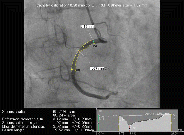
Fig. 5.2
Example of quantitative stenosis assessment by quantitative coronary angiography (QCA). Conventional coronary angiography (CCA) image of the right coronary artery (RCA) is shown demonstrating longitudinal measurements of percentage diameter stenosis, calculated by comparing minimal luminal diameter at the site of maximal stenosis (C) with normal reference diameters proximal (A) and/or distal (B). Percentage diameter stenosis = {1 − C/[(A + B)/2]} × 100 (%)
Manually or semiautomatically
Drawing of diameter using cross-sectional or longitudinal lumen display with similar accuracy compared with QCA
Advantage of cross-sectional displaying: assessing all lumen borders in 1 view
One important caveat for quantitative stenosis assessment by MDCT: adjustment of window width and level settings
A trend to establish quantitative method by MDCT evaluation as the routine assessment also for clinical applications
Reporting by 2° (commonly used reporting method in coronary CTA):
Insignificant: 0–50 % diameter stenosis
Significant: 51–99 % diameter stenosis
Table 5.2
Recommended qualitative and quantitative stenosis grading by the Society of Cardiovascular Computed Tomography (SCCT)
Descriptive lumen obstruction | Qualitative stenosis grading | Quantitative stenosis grading |
|---|---|---|
Normal | Absence of plaque/no luminal stenosis | Absence of plaque/no luminal stenosis |
Minimal | Plaque with negligible impact on lumen | Plaque with <25 % stenosis < div class='tao-gold-member'>
Only gold members can continue reading. Log In or Register to continue
Stay updated, free articles. Join our Telegram channel
Full access? Get Clinical Tree
 Get Clinical Tree app for offline access

|