Assessment of Perioperative Events and Problems

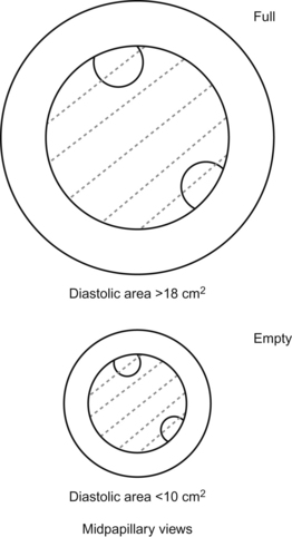
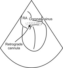
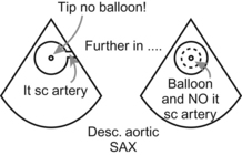
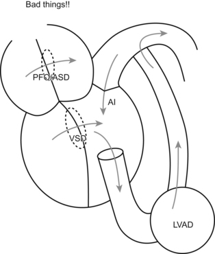
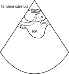
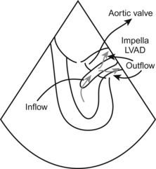
Chapter 16


Assessment of Perioperative Events and Problems


Ricardo Martinez-Ruiz and Christopher J. Gallagher


Hypotension and Causes of Cardiovascular Instability


The TEE is the anticrash weapon of choice.


When badness happens (and we’ve all seen it happen), you might not have a Swan or CVP. And even if you do, you’re still wrestling with numbers that tell you something, but not the whole picture. You are left with a set of numbers from which you infer, or hope, you have the picture.


The TEE gives you the real picture, right now, no need for a leap of faith.


Cahalan points out in the tapes and at the meetings that, even with only a little TEE experience, most people can diagnose the most common problems in mere minutes. After all, when most patients go to caca, you want to know:


For most problems, then, Cahalan gives us a nice, neat, easy-to-understand and inherently obvious breakdown of the main causes of hypotension:


1. End-diastolic area decreased, ventricle contracting OK—You’re low on volume. The heart is empty, so fill it.


2. End-diastolic area increased, ventricle contracting poorly—You have a bad and already overfilled ventricle. Fix whatever’s causing the global hypokinesis (Get a blood gas! Don’t forget the basic stuff!), and once you’ve fixed what you can fix, it’s time for inotropes or ventricular support of some kind.


3. End-diastolic volume normal or low, ventricle contracting well or hyperdynamic—You have a problem with the volume not “going where it’s supposed to go”. Either the volume is all going out into a vastly dilated circulatory tree (anaphylaxis with low systemic vascular resistance), or else some other channel is misrouting your good cardiac output (severe mitral regurg or aortic regurg, or a ventricular septal defect).


image


The first two are easy to see with a glance at the TEE. The third is a little trickier, but you can augment your TEE findings with other stuff. (Flushed appearance and wheezing going along with anaphylaxis; murmurs or further TEE views to find mitral regurg, aortic regurg, or a VSD.)


Then the final thing you want to know, “Tamponade, yes or no?”, is figure-outable with your basic search for a pericardial effusion plus the hemodynamics of tamponade.


image


If you take nothing else away from TEE (say you don’t want to bother taking the TEE exam), if you at least know this, the differential for hypotension, you will save somebody some day.


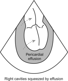
Cardiac Surgery: Techniques and Problems


Assessment of Bypass and Cardioplegia


How the hell do you use TEE to assess bypass and cardioplegia? Got me. I have no clue what the Society of Cardiovascular Anesthesia folk were thinking when they put this on their magical list.


Let’s stretch a little and try to figure this one out.


One thing is worth mentioning at this time. Disconnect your echo probe while on bypass. That will allow the probe to cool down and prevent esophageal burns. And remember that you do need to disconnect the probe, not just put the image on FREEZE. Although the word FREEZE implies a cool state of affairs, you have just frozen the image. You haven’t actually frozen the probe and turned it into a big Fudgecicle.


Cannulas and Devices Commonly Used During Cardiac Surgery


The main cannula you’ll be asked to visualize is the retrograde cannula for cardioplegia. First, you need to know where the coronary sinus is (the site of the retrograde cannula).


To see the coronary sinus on TEE, you need to get your ME 4-chamber view and slowly advance the probe into the stomach and VOILA!!! The coronary sinus shows up as a drak conduit that drains into the right atrium (you can check with Doppler to see the coronary sinus blood flow). Ocassionally you may have a valve (Thebesian valve) impeding the easy positioning of the retrograde cannula, the cannula gets stuck on it. It is pretty cool to tell your surgeon that the reason he is struggling getting it in position is because of the valve.


image


Cannula to cross the threshold. Like all cannulas, the retrograde cannula has a “double line” that tells you it’s a man-made thingamajig. And like all cannulas, its 3-D reality will dive in and out of your 2-D picture, making it sometimes a little tough to keep it entirely in view. In some occasions you may see the stippled balloon at the end of the cannula: it looks like a star cluster (yes…use your imagination!).


Circulatory Assist Devices


It’s not a stretch to look for correct placement of an intra-aortic balloon pump. You want to see the tip of the IABP at the takeoff to the left subclavian artery. No higher (occlusion to the left arm) and no lower (inadequate function of the balloon pump and potential occlusion of renal arteries, oops). You should see the tip of the IABP, but no balloon!, at the level of the takeoff of the Lt subclavian artery on the most upper view of the descending thoracic aorta. As soon as you push the probe deeper, the balloon should be visible with its characteristic up and down, up and down… (you can also assess the quality of expansion of the balloon).


image


Things can get more exotic, of course.


Left and right ventricular assist devices, and ECMO when things are going really swell, all enter the cardiac realm in this Brave New World we inhabit.


With an LVAD, you want to make sure the person doesn’t have a patent foramen ovale or interatrial septal defect. You could, as the blood rushes out of the left side into the assist device, “suck” blood from the right side over to the left.


If this happens, then no blood goes out the right side, so no blood goes to the lungs, so no oxygen enters the body. Unless your patient is a cyanobacterium, he or she will need oxygen.


So check for these PFO when you start an LVAD. You may also need to make sure that there is no aortic regurgitation. If that happens blood will “recirculate” in a circle of death >>>> from the LV >>> to LVAD >>>> to aorta >>> back to LV.


image


Once the LVAD is going, you can also use the TEE to confirm that the aortic valve isn’t opening. At first that concept seems a little jarring.


Yes, usually that is the case, but remember, you’re not in Kansas anymore, Dorothy. The LVAD is doing all the work now. You want all the blood to leave the heart and go into the machine.


In testville, remember that the cannula that is draining blood out of the body into the machine is the inflow cannula. (That is, inflow as far as the machine is concerned. You could get faked out and think, well, relative to the body, that is technically outflow, so… Don’t think that!)


There are two new gadgets for LV support that can be placed percutaneously >> the Tandem heart and the Impella. The Tandem inflow cannula goes from the femoral vein into the IVC into the RA and then goes through the interatrial septum into the LA! The LA blood is pulled into the centrifugal pump located outside the body to be reinfused into the arterial system via the femoral artery to provide systemic perfusion. You can check the placement of the inflow cannula as it goes across the interatrial septum.


image


The impella is different: it consists of a very small impeller-based pump that its placed across the aortic valve (via the femoral artery most commonly) and sucks blood out of the LV and propels it back into the aorta. This will provide for overall systemic perfusion needs and will also let the LV decompress and recover. Neat stuff!!!


image


ECMO and RVADs? You can use TEE to check those cannulas too and just make sure they seem to be in the right place.


Jun 4, 2016 | Posted by in CARDIOLOGY | Comments Off on Assessment of Perioperative Events and Problems

Full access? Get Clinical Tree

Get Clinical Tree app for offline access