Assessment of Ischemic Heart Disease Utilizing Dobutamine


Assessment of Ischemic Heart Disease Utilizing Dobutamine


Rolf Gebker

Andreas Schuster

Eike Nagel


INTRODUCTION

Ischemic heart disease is one of the most common health problems of the Western world. A variety of tests are available in routine clinical practice for the noninvasive diagnosis of coronary artery disease (CAD) such as exercise electrocardiography (ECG), echocardiography, single photon emission computed tomography (SPECT), positron emission tomography (PET), and cardiovascular magnetic resonance (CVMR) imaging. The advantages and disadvantages of each technique are summarized in Table 16.1. Many noninvasive diagnostic tools are suboptimal and both patients and physicians want a reliable diagnosis. Consequently, 40% to 60% of all patients who undergo invasive cardiac catheterization procedures do not require a revascularization procedure such as bypass surgery or angioplasty. Even more importantly, it has been shown that revascularization procedures in patients without flow-limiting coronary artery stenoses result in a higher event rate than optimal medical therapy alone (1,2). As a consequence, current guidelines have stressed the importance of documenting ischemia, preferably using noninvasive functional testing before elective invasive procedures (3). Thus, a noninvasive test with a high rate of diagnostic accuracy is required to optimally guide patient management and avoid unnecessary invasive tests or potentially harmful procedures.

CVMR has evolved into a new technique for the noninvasive detection of obstructive CAD. The ability of CVMR to visualize global and regional wall motion and systolic thickening of the left ventricle (LV) with a high degree of spatial and temporal resolutions makes it possible to detect even subtle abnormalities of wall motion. In addition, perfusion defects and reductions in coronary flow reserve can be assessed. Except for high-grade coronary artery stenoses, abnormalities can, for the most part, be identified only under stress conditions. These can be induced by physical exercise or by means of standardized stress protocols with infusions of pharmacologic agents such as dobutamine/atropine, dipyridamole, or adenosine. To date, the most reliable clinical data are either based on the analysis of LV wall motion and thickening during dobutamine stress or on the analysis of first pass perfusion during adenosine stress. In this chapter, the evidence and a detailed description of how to utilize dobutamine as a pharmacologic agent to assess both viability and ischemia are presented.


HOW TO PERFORM DOBUTAMINE STRESS TESTING

Stress testing can be performed with either physical or pharmacologic stimulation. In general, to detect ischemic heart disease with a high level of sensitivity and reproducibility, a defined endpoint (submaximal stress) must be reached. This endpoint is defined by the heart rate as follows:

Target heart rate = 0.85(220 – Age)


ERGOMETRIC STRESS

Ergometric stress is the most physiologic stress test and thus generally the preferred method for testing. Patients exercise on a bicycle or treadmill ergometer with incremental workloads.

Despite the fact that ergometric stress in combination with CVMR has been shown to be feasible both for using a magnetic resonance (MR)-compatible bicycle while lying on the examination table or a treadmill placed outside the 5-Gauss line inside the MRI room, there are several limitations associated with these techniques (4,5). Patients have to stop exercising and must be moved back into the bore for scanning, which may cause the heart rate to fall below the target heart rate thus reducing sensitivity. Due to physical exercise, breathing is generally more labored and breathholding more challenging, which may introduce motion artifacts. Most importantly; however, patients are often not able to reach the anticipated target because of comorbidities such as peripheral vascular or degenerous musculoskeletal disease or due to chronic deconditioning. As a result, a large number of tests may be nondiagnostic because of an inadequate level of stress or image quality. Thus, the most robust approach for stress testing within the CVMR environment is pharmacologic stress testing.


PHARMACOLOGIC STRESS

Pharmacologic stress is preferred by many clinicians because the results are highly reproducible and diagnostic in most patients. Careful monitoring is required for patient safety. Two different approaches are used: Oxygen consumption can be increased by increasing the heart rate and contractility (dobutamine), or vasodilation with the induction of regional flow heterogeneities (steal effect) can be induced (dipyridamole/adenosine). The different pharmacologic stress protocols are summarized in Table 16.2.

Dobutamine is a sympathomimetic drug with β1-, β2-, and slight α1-receptor-stimulating properties. Infusion of the drug increases the cardiac contractility and rate and decreases the systolic vascular resistance. During low-dose infusion (≤10 µg/kg/min), an increase in contractility is the major effect, whereas at higher doses, the augmented consumption of oxygen causes contraction abnormalities in myocardial segments supplied by stenotic coronary arteries.

A low dose of dobutamine is defined as an infusion of up to 10 µg/min/kg of body weight. Such a dose is sufficient to stimulate myocardium that does not contract at rest but may benefit from revascularization (viable or hibernating myocardium); however, this dose is insufficient to induce ischemia.

A high dose of dobutamine is defined as an infusion aimed at inducing ischemia. As previously explained, it is essential that patients reach their target heart rate. To achieve this goal, atropine, which increases the heart rate by an
anticholinergic mechanism, is commonly added. The stress protocol most widely used is described in the guidelines of the Society of Cardiovascular Magnetic Resonance (6). Stress is induced by increasing doses of dobutamine, started at 10 µg/kg of body weight per minute for 3 minutes and increased in increments of 10 µg/kg of body weight per minute every 3 minutes, until a maximal dose of 40 µg/kg of body weight per minute is reached. (Some investigators use 50 µg/kg of body weight per minute as a maximum.) If the target heart rate is not reached, up to 2 mg of atropine is added in 0.25-mg fractions. The test must be stopped if certain criteria are fulfilled (Table 16.3).


TABLE 16.2 Stress Protocols


Stress Test


Patient Instructions


Protocol


Antidote


Dobutamine for the assessment of viability


5, 10 µg/kg BW per minute for >3 min


Dobutamine/atropine for the detection of myocardial ischemia (wall motion and perfusion)


No β-blockers and nitrates 24 h before the examination


5, 10, 20, 30, 40 µg/kg BW per minute for 3 min each, up to 2 mg atropine (8 × 0.25 mg) until submaximal heart rate [(220 – age) × 0.85] is reached (half-life 2 min)


β-blocker (esmolol) 0.5 mg/kg as slowly injected bolus, additional bolus of 0.2 mg/kg as needed, sublingual nitroglycerin


Dipyridamole (perfusion)


No caffeine (tea, coffee, aminophylline 250 mg chocolate, etc.) or medications such as aminophylline or nitrates 24 h before the examination


0.56 mg/kg BW per minute for 4 min, maximal effect after approximately 3-4 min (halflife 30 min)


Aminophylline 250 mg IV slowly injected with ECG monitoring, sublingual nitroglycerin


Adenosine (perfusion)


Same as for dipyridamole


140-210 µg/kg BW per minute for 3-6 min (half-life 4-10 s)


Stop infusion (in occasional cases aminophylline 250 mg IV slowly injected with ECG monitoring)


BW, body weight; ECG, electrocardiogram.


IMAGE ACQUISITION

For the assessment of wall motion, cine loops of the heart are acquired with steady state free precession (SSFP) usually in combination with parallel image acquisition and retrospective ECG gating, resulting in 25 to 50 phases per cardiac cycle up to heart rates of 200 beats per minute during a breath-hold of 6 to 10 seconds. In contrast to turbo gradient-echo techniques that have been used in older studies, imaging contrast in SSFP results in a much better delineation of the endocardial border, especially in long-axis views (7). The in-plane spatial resolution is in the range of 1.6 × 1.6 mm with a slice thickness of 6 to 8 mm. The heart can be visualized with either contiguous short-axis slices or a combination of several short-axis (typically basal, mid, and apical slice) and long-axis (typically two-, three-, and four-chamber) views (Fig. 16.1).


TABLE 16.3 Dobutamine Termination Criteria


Submaximal heart rate reached [(220 — age) × 0.85]


Systolic pressure decrease >20 mm Hg below baseline systolic blood pressure or decrease >40 mm Hg from a previous level


Blood pressure increase >240/120 mm Hg


Intractable symptoms


New or worsening wall-motion abnormalities in at least 2 of 17 adjacent left ventricular segments


Complex cardiac arrhythmias


Imaging is performed at rest and then immediately after the dobutamine dose is increased for each stress level. Images are acquired and reviewed immediately at rest and during each stress level to detect new wall-motion abnormalities during stress. The stress protocol, details of monitoring, contraindications, and termination criteria are summarized in Tables 16.2,16.3,16.4 and 16.5. Dobutamine stress magnetic resonance (DSMR) is generally performed on 1.5-T scanners. With the advent of 3 T for cardiac imaging, there has been a growing interest to perform high-dose dobutamine stress protocols under these high field conditions. However, this approach has been hampered by an increase in artifacts (banding, flow-related artifacts), especially applying SSFP imaging during higher heart rates. Turbo gradient-echo cine imaging with administration of contrast agent offers more robust imaging under these conditions (8). More recently, the development of dual-source RF transmission with patient-adaptive local RF shimming has led to an improvement in SSFP image quality during DSMR at 3 T (9).


SAFETY

During stress examinations in which low or high doses of dobutamine are administered, monitoring the patient within the magnet is mandatory. In general, monitoring during a CVMR examination requires that the same precautions be taken and the same emergency equipment be available as in any other stress examination. A physician trained in cardiovascular emergencies and resuscitation must be at the
scanner. Apart from specific contraindications to CVMR such as the presence of retro-orbital metal, cerebral clips, or non-MRI compatible pacemakers, the contraindications are identical to those for stress echocardiography and are listed in Table 16.4.


Figure 16.1. DSMR in a patient with new onset angina after coronary bypass grafting and resting wallmotion abnormalities of the inferolateral basal wall. During high-dose dobutamine stress, the patient develops an extensive stress-inducible wall-motion abnormality of the anterior/anterolateral wall from the base to the apex (white arrows). Invasive coronary angiography demonstrated extensive coronary disease with a highgrade stenosis of the LAD distal to the anastomosis of the LIMA-graft (black arrow). SAX, short axis views; ED, end diastolic frame; ES, end systolic frame.

In a routine clinical setting, DSMR proved to be feasible and safe, and it resulted in a high number of diagnostic ex aminations (89.5%) in patients without contraindications to MRI (10). Wahl et al. (10) reported a 5-year experience of high-dose DSMR in 1,000 consecutive patients and showed a safety profile virtually identical to that previously reported for dobutamine stress echocardiography. One patient suffered sustained ventricular tachycardia and emergent defibrillation was carried out, while no cases of death or myocardial in farction occurred over the years (Table 16.6). Approximately 50% of the patients had inducible ischemia during stress CVMR, thereby closely reflecting the clinical practice in a tertiary center. Data from the large Euro CVMR registry has
confirmed the low periprocedural complication rate of highdose DSMR (11). Thus, although adverse events are rare, the staff must be prepared to remove the patient from the magnet rapidly if necessary and must comply closely with the test termination criteria (
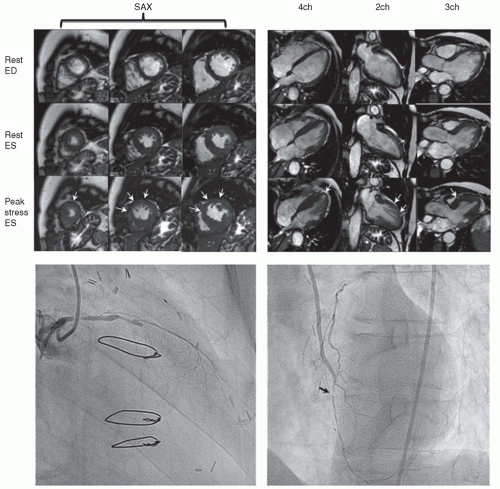
Table 16.3). Whereas in most other modalities, “eye-to-eye” contact between patient and examiner takes place, communication during CVMR is usually established via a microphone system and video cameras, although it can also be conducted personally. This does not hinder the safety process if the patient is monitored carefully for symptoms, blood pressure changes, and wall-motion abnormalities (Table 16.2). Either standard equipment can be placed outside the scanner room and connected to the patient with special extensions through a waveguide in the radiofrequency cage or special CVMR-compatible equipment, available at many CVMR sites, can be used. A defibrillator and all medications needed for emergency treatment must be available at the CVMR site. A specific problem associated with monitoring within the magnet is that of assessing ST-segment changes from the ECG. However, because wallmotion abnormalities precede ST changes and because such abnormalities can be readily detected with fast CVMR, monitoring is effective without a diagnostic ECG and can also be performed in patients with left-bundle branch block, who are routinely evaluated with dobutamine stress echocardiography despite nondiagnostic ST segments. Such wall-motion abnormalities can be detected immediately after image reconstruction, which is completed 5 to 10 seconds after image acquisition. In addition, real-time imaging permits the immediate detection of wall-motion abnormalities and can be used for monitoring (12). Such real-time acquisition has been found to yield a similar diagnostic accuracy for the detection of inducible wall-motion abnormalities (sensitivity and specificity for real-time MR imaging 81% and 83%) when compared to conventional MR cine imaging (13). However, spatial resolution is less, and thus we recommend ECG-triggered breathhold imaging as the basis for the final diagnosis.


TABLE 16.4 Contraindications to Magnetic Resonance Stress Tests


MR examination


Incompatible metallic implants (e.g., pacemakers, retro-orbital metal, cerebral artery clips)


Claustrophobia


Dobutamine


Severe arterial hypertension (>22 0/120 mm Hg)


Unstable angina pectoris


Significant aortic stenosis (aortic valve gradient >50 mm Hg or aortic valve area >1 cm2)


Complex cardiac arrhythmias


Significant hypertrophic obstructive cardiomyopathy


Myocarditis, endocarditis, pericarditis


Other major disease


Dipyridamole/adenosine


Myocardial infarction <3 d


Unstable angina pectoris


Severe arterial hypertension


Asthma or severe obstructive pulmonary disease


Atrioventricular block >Iia


TABLE 16.5 Monitoring Requirements for Stress Magnetic Resonance Imaging


Dobutamine


Adenosine


Heart rate and rhythm (single-lead ECG)


Continuously


Continuously


Blood pressure


Every minute


Every minute


Pulse oximetrya


Continuously


Continuously


Symptoms


Continuously


Continuously


Wall-motion abnormalities


Every dose increment


Not necessary


a Only required for additional rhythm control if no vector ECG is available.


ECG, electrocardiogram.


TABLE 16.6 Moderate and Severe Side Effects in 1,000 Patients, Including Positive Tests


Sustained VT


1 (0.1%)


Nonsustained VT


4 (0.4%)


Paroxysmal atrial fibrillation


16 (1.6%)


Temporary AV block II 2:1


2 (0.2%)


RR-increase (>240/120)


5 (0.5%)


RR-decrease >40 mm Hg


5 (0.5%)


Nausea


31 (3.1%)


Total


64 (6.4%)


With permission from: Wahl A, Paetsch I, Gollesch A, et al. Safety and feasibility of high-dose dobutamine-atropine stress cardiovascular magnetic resonance for diagnosis of myocardial ischaemia: Experience in 1000 consecutive cases. Eur Heart J 2004;25(14): 1230-1236.


IMAGE INTERPRETATION

For image interpretation, a multiple cine loop display that allows different stress levels to be assessed at the same time is essential. The ventricle is typically analyzed for 17 LV segments per stress level according to the standards suggested by the Society of Cardiovascular Magnetic Resonance (6) and the American Heart Association (Fig. 16.2). Each segment is assigned to a specific coronary artery; however, depending on the coronary artery anatomy or degree of collateralization, some segments may be supplied by different arteries. Thus, it is sometimes not possible to define a stenotic coronary artery from a wall-motion study. Image quality is graded as excellent, good, moderate, or nondiagnostic, and the number of diagnostic segments is reported. Segmental wall motion is classified as normokinetic, hypokinetic, akinetic, or dyskinetic and assigned one
to four points (Fig. 16.3). The sum of the points is divided by the number of analyzed segments to yield a wall-motion score. Normal contraction results in a wall-motion score of one, and a higher score indicates wall-motion abnormality. During stress with increasing doses of dobutamine, either a lack of increase in wall motion or systolic wall thickening, or a reduction in wall motion or thickening is regarded as a pathologic finding (Fig. 16.1).


Figure 16.2. Seventeen-segment model suggested by the American Heart Association. The coronary artery territories are shown in the graph.


ACCURACY OF STRESS-INDUCED WALLMOTION ABNORMALITIES IN THE DIAGNOSIS OF ISCHEMIA

The echocardiographic detection of wall-motion abnormalities during high-dose dobutamine or exercise stress has been shown to be an accurate diagnostic tool for screening patients with suspected CAD. Sensitivities of 54% to 96% and specificities of 60% to 100% have been reported (14), depending on the pretest likelihood of disease and the experience of the stress centers. However, the value of stress echocardiography is limited by a 10% to 15% rate of nondiagnostic results (14) and low specificities for the basal-lateral and basal-inferior segments of the LV (15).

The first studies applying dobutamine as a stress agent in the CVMR environment have yielded good results in the detection of wall-motion abnormalities at intermediate doses of dobutamine (maximum of 20 µg/kg of body weight per minute intravenously) (Table 16.7) (16,17,36

Only gold members can continue reading. Log In or Register to continue

Stay updated, free articles. Join our Telegram channel

May 24, 2016 | Posted by in CARDIOLOGY | Comments Off on Assessment of Ischemic Heart Disease Utilizing Dobutamine

Full access? Get Clinical Tree

Get Clinical Tree app for offline access