Assessment of Congenital Heart Disease in the Adult Patient


Assessment of Congenital Heart Disease in the Adult Patient


Isobel Russell

Elyse Foster


This chapter is, in large part, based on an original review that is currently in press: Russell IA, Rouine-Rapp K, Stratmann G, Miller-Hance W. Congenital Heart Disease in the Adult: A Review with Internet-Accessible Transesophageal Echocardiography, Anesthesia and Analgesia.

Advances in cardiac surgery, anesthesia, intensive care, and diagnosis over the last 50 years have permitted survival of 85% of infants with congenital heart disease (CHD) into adulthood. In fact, for the first time, the number of adults with CHD equals the number of children with the disorder (1). Although accurate statistics are lacking, the number of adults with CHD in the United States has reached a conservative estimate of approximately 800,000, which is increasing at the rate of 5% per year. It was estimated that by the year 2002, the number of adults with simple, moderate, and complex forms of CHD would be approximately 370,000, 300,000, and 120,000, respectively (1). These figures are underestimates because they do not include patients that are diagnosed in adulthood, but are based on the widely varying reported prevalence rates of congenital heart disease. It is estimated that the number of patients with congenital heart disease reaching adulthood is approximately 16,000 per year in the United States alone (1). The recent consensus report (1) suggests that these patients should receive care in regional centers for adult congenital heart disease and the patients should keep their own copies of information on operations, cardiac catheterizations, and other diagnostic tests, such as echocardiograms in the form of a “health passport.”


INDICATIONS

Indications for TEE in adult patients with congenital heart disease should follow established guidelines by the American Society of Anesthesiologists and the Society of Cardiovascular Anesthesiologists (2) and the American College of Cardiology/American Heart Association Task Forces (3). It should be emphasized that a complete diagnosis of the congenital heart lesion should be defined prior to performance of intraoperative TEE. The lesion, coexisting pathology, hemodynamics, cardiac size, and function should be well delineated whether by cardiac catheterization, transthoracic echocardiography, or transesophageal echocardiography (4,5,6). Intraoperative TEE should not be used as a replacement for an inadequate preoperative evaluation.

Here are summaries from the two reports on the indications for intraoperative TEE for congenital heart disease.

a) “Practice guidelines for perioperative transesophageal echocardiography.”

Most cardiac defects requiring repair under cardiopulmonary bypass are a category 1 indication for intraoperative TEE, including pre- and post-cardiopulmonary imaging (category 1 is defined as that being supported by the strongest evidence or expert opinion substantiating that TEE is useful in improving clinical outcomes) (2).

b) “ACC/AHA guidelines for the clinical application of echocardiography.”

Monitoring and guidance during cardiothoracic procedures associated with the potential for residual shunts, valvular regurgitation, obstruction or myocardial dysfunction is a class 1 indication (defined as conditions for which there is evidence and/or general agreement that a given procedure or treatment is useful and effective) (3). This was defined under the indications for TEE in pediatric patients with congenital heart disease but is applicable to adolescents and adults with CHD.


Approach

Image orientation, nomenclature guidelines, and a comprehensive intraoperative echocardiographic examination
should follow those suggested by the American Society of Echocardiography (7) and ASE/SCA guidelines (8). Appropriate TEE views for evaluation of congenital heart lesions are described in Table 20.1 and Fig. 20.1. Assessment of the connections of the various cardiac segments, atrial arrangement or situs, venoatrial, atrioventricular, and ventriculoarterial connections should be performed. Septal and valvar structures should then be evaluated, including assessment of flow velocities with Doppler echocardiography. In general, the approach for each lesion is to define the intracardiac anatomy and associated defects, assess the ventricular function, and evaluate any residual lesions (Table 20.2). Comprehensive reference atlases include Pediatric Echocardiography, edited by N. Silverman (Williams and Wilkins), Transesophageal Echocardiography in Congenital Heart Disease, edited by O. Stumper and G. Sutherland (Little, Brown and Company), and Congenital Heart Disease in Adults, edited by J. K. Perloff and J. S. Child (W. B. Saunders).


TABLE 20.1. Congenital Heart Lesions—Appropriate TEE Views for Evaluation of Congenital Heart Lesions


Lesion


TEE Plane


Atrial Septal Defects


ME four-chamber for ostium secundum and primum defects


ME bicaval for interatrial septum for sinus venosus defects and possible anomalous pulmonary veins


Ventricular Septal Defects


ME four-chamber, LAX for perimembranous, inlet and muscular VSDs, chamber sizes, presence of ventricular septal aneurysm


ME AV LAX, deep TG LAX views for evaluation of the aortic valve for insufficiency and herniation


ME RV inflow and outflow for pulmonic valve insufficiency


Atrioventricular Septal Defects


ME four- and two- chamber for the bridging leaflets and their attachments, extent of intracardiac defects, size of septal defects, and extent of AV valve regurgitation


Aortic Stenosis (valvular and subvalvular)


Deep TG LAX for aortic stenosis, insufficiency, and aortic root size


ME AV SAX evaluation of aortic valve morphology


ME four-chamber for LV hypertrophy and function assessment


Transposition of the Great Arteries


ME four-chamber to evaluate AV valve regurgitation and baffle assessment after Senning/Mustard procedures


ME bicaval view additionally for caval junctions and pulmonary veins


TG mid SAX for ventricular function and SWMA


Deep TG LAX for ventriculoarterial connections and anastomas after arterial switch


Tetralogy of Fallot


ME AV LAX and deep TG LAX for definition of aortic override and obstruction of the right ventricular outflow tract and estimation of gradients


ME RV inflow-outflow for RVOT evaluation


ME four-chamber view for position and extension of VSD and other additional VSDs


Truncus Arteriosus


ME four-chamber view for VSD position and extent


ME AV SAX to evaluate truncal valve


ME AV LAX and deep TG LAX for evaluation of truncal insufficiency and determination of truncal anatomy


Patent Ductus Arteriosus


Difficult to visualize by 2D TEE, but ductal flow can be visualized in the ME asc aortic SAX view by presence of abnormal continuous high velocity aliased flow


Pulmonic Valve Stenosis


ME RV inflow-outflow and deep TG LAX view for outflow tract evaluation and gradient estimation


ME asc aortic SAX for evaluation of pulmonic valve and main pulmonary artery


Single Ventricles


ME four-chamber, two-chamber and deep TG LAX for atrioventricular morphology and atrioventricular and ventriculoarterial connections


ME bicaval view for evalution of Glenn anastomosis


asc, ascending; AV, aortic valve; LAX, longitudinal axis; ME, midesophageal; RV, right ventricle; RVOT, right ventricular outflow tract; SAX, short axis; SWMA, segmental wall motion abnormality; TG, transgastric.


Congenital heart disease can be classified in several ways: according to the underlying physiology of either shunt; obstructive, regurgitant, and mixed lesions; according to the presence and absence of cyanosis; or according to the level of complexity of the lesion. For purposes of simplicity, this chapter will organize the lesions according to level of complexity, with greater emphasis on the more common lesions.


SIMPLE LESIONS


Atrial Septal Defects

Atrial septal defects (ASD) are one of the most common defects in the adult population, accounting for one-fourth
to one-third of all lesions, occurring more commonly in women (9). There are four types of atrial septal defects:


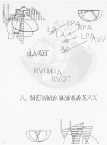
FIGURE 20.1. Cross-sectional views depicting imaging planes useful for assessment of congenital heart defects. The appropriate multiplane angle is indicated by the icon on the right adjacent to each view. The imaging plane is indicated by the schematic drawing of the heart on the left. asc., ascending; Ao, aorta; AoV, aortic valve; IVC, inferior vena cava; LA, left atrium; LAA, left atrial appendage; LAX, long axis; LPA, left pulmonary artery; LV, left ventricle; LVOT, left ventricular outflow tract; ME, midesophageal; MPA, main pulmonary artery; RA, right atrium; RPA, right pulmonary artery; RV, right ventricle; RVOT, right ventricular outflow tract; SAX, short axis; SVC, superior vena cava; TG, transgastric; TV, tricuspid valve.


TABLE 20.2. Indications for Primary Surgery, Common Postoperative Complications and Indications for Reoperation in Congenital Heart Disease


Lesion


Indication for Primary Repair


Post-Repair Complications


Indication for Reoperation


Atrial Septal Defect


Qp:Qs ≥1.8:1 Paradoxical embolization


Atrial fibrillation, CVA*


Secundum: none Primum: MR


Ventricular Septal Defect


Qp:Qs ≥ 1.5:1


Patch leak Endocarditis* Progressive PHTN


Patch leak when Qp:Qs ≥ 1.5:1


Patent Ductus Arteriosus


Qp:Qs ≥ 1.5:1


Persistent shunt Endoarteritis* Progressive PHTN


Persistent shunt


Atrioventricular Canal Defect


Qp:Qs > 1.5:1 Mitral regurgitation


Patch leak Endocarditis Progressive MR Atrial fibrillation Atrioventricular block


MR Patch leak


Anomalous Pulmonary Venous Drainage


Qp:Qs > 1.5:1


Obstruction of the pulmonary veins


N/A


Tetralogy of Fallot


No previous repair


Patch Leak Pulmonary insufficiency Residual PS Heart block Ventricular and atrial arrhythmias Sudden death Endocarditis


Hemodynamically significant PI or PS


Transposition of Great Vessel


N/A


Postatrial Switch: RV failure Tricuspid regurgitation Baffle obstruction Atrial arrhythmias Heart block Endocarditis


Postatrial Switch: Supravalvar aortic stenosis (neopulmonic stenosis)


Coronary artery stenosis


Significant baffle obstruction Progressive RV failure Tricuspid regurgitation


Ebstein’s Anomaly


Severe tricuspid regurgitation RV failure


Atrial arrhythmias Progressive TR


Severe TR


Tricuspid Atresia


N/A


Post-Fontan Atrial arrhythmias Protein losing enteropathy Ascites


Conduit obstruction


Aortic Stenosis


AVA < 0.8 cm2 in presence of symptoms


Post-Valvotomy


Progressive AI or restenosis Ventricular arrhythmias Sudden death Endocarditis


Severe AS or AI in the presence of symptoms or LV dysfunction


Subaortic Stenosis


Gradient > 30 mm Hg or the development of AI at a lower gradient


Progressive AI Restenosis Endocarditis


Severe AI or recurrent stenosis in the presence of symptoms


Pulmonary Stenosis


Transpulmonary gradient > 50 mm Hg


Transpulmonary gradient < 50 mm Hg with RV hypertrophy or symptoms


Restenosis Pulmonic valve insufficiency Endocarditis rare


Severe PS or PI in the presence of symptoms


Coarctation of Aorta


Upper extremity HTN


Transcoarct gradient > 25 mm Hg


Residual HTN Saccular aneurysm Dissecting aneurysm Circle of Willis aneurysm CVA Premature CAD AS/AI Endocarditis or endarteritis


Recurrent coarctation AS/AI (see above) CAD Saccular or dissecting aneurysm


Qp:Qs, pulmonary to systemic flow ratio; CVA, cerebrovascular accident; PHTN, pulmonary hypertension; MR, mitral regurgitation; PI, pulmonic valve insufficiency; PS, pulmonary stenosis; AS, aortic stenosis; TR, tricuspid regurgitation, RV, right ventricle; AVA, aortic valve area; CAD, coronary artery disease.


* These complications are most common when repair performed at age > 40; *endocarditis and endarteritis likely only in the presence of persistent shunt.


1. Ostium secundum defect (70%), which occurs in the midseptum region of the fossa ovalis. Varying degrees of mitral valve prolapse and mitral regurgitation can occur, but hemodynamically significant lesions are uncommon.

2. Ostium primum defect (15%-25%), which is also a form of a partial atrioventricular canal defect and consists of an ASD in the lower part of the interatrial septum. Abnormalities of the atrioventricular valve can occur with a “cleft” anterior mitral valve and septal tricuspid valve leaflet with variable degrees of regurgitation.

3. Sinus venosus defect (10%) is usually superior and posterior in relation to the superior vena cava (more frequently) and/or inferior vena cava. These defects are frequently associated with an anomalous drainage of one or more pulmonary veins into the right atrium or superior vena cava (10).

4. Coronary sinus defects (extremely rare) occur between the left atrium and coronary sinus and can be associated with a persistent left superior vena cava.

Imaging with intraoperative TEE is performed following the guidelines in Table 20.1 to confirm the presence, size, and location of the defect; degree of atrioventricular valve regurgitation; ventricular function; and associated anomalies, such as anomalous pulmonary veins (Fig. 20.2). Postoperatively, TEE is used to detect residual shunts by color Doppler and contrast, assess ventricular function, and assess for pulmonary venous obstruction and valvar regurgitation. To evaluate for residual shunts, we use agitated saline contrast because the microbubbles are readily apparent even when a very small number of microbubbles cross a defect (11). The method used is to vigorously agitate 0.5-1.0 cc of the patient’s blood between two syringes with 10 cc of saline (12).

A defect in the interatrial septum allows pulmonary venous return to pass from the left to the right atrium. Because this left-to-right shunt increases the venous return to the right ventricle, the right ventricular stroke volume and pulmonary blood flow are increased compared with the systemic blood flow. Right ventricular volume overload results. Indirect evidence of an atrial septal defect includes right-sided chamber enlargement and appearance of saline contrast in the left heart chambers. Color flow Doppler echocardiography can demonstrate flow across the atrial septum and detect mitral or tricuspid regurgitation.

Primary or patch closure of an atrial septal defect in childhood provides excellent operative results and nearly normal long-term survival in adults (13,14). Additionally, a recent retrospective study suggested improved 10-year survival in patients over the age of 40 years treated surgically (95%) compared with those treated medically (84%) (14). A prospective clinical trial (15) randomized adult patients with secundum atrial septal defects with shunt ratios > 1.7:1 to surgical versus medical management, showing improved survival with surgical closure. However, late repair does not appear to reduce the incidence
of arrhythmias, which are generally related to preoperative atrial dilatation or postoperative incisional reentry (16). Operative patch closure is generally recommended if the shunt is large with a pulmonary blood flow-to-systemic blood flow ratio of 1.8:1 or higher and right ventricular enlargement. Percutaneous closure with a variety of devices is becoming increasingly available and can be considered in adults. Patients with smaller shunts have a lower incidence of congestive heart failure, pulmonary hypertension, and arrhythmias but are at risk for paradoxical embolization. In patients with ostium primum defects, surgical valve repair with or without annuloplasty may reduce the severity of the mitral and tricuspid regurgitation. If severe mitral regurgitation persists, valve re-repair or replacement is necessary.

Only gold members can continue reading. Log In or Register to continue

Stay updated, free articles. Join our Telegram channel

Jul 15, 2016 | Posted by in CARDIOLOGY | Comments Off on Assessment of Congenital Heart Disease in the Adult Patient

Full access? Get Clinical Tree

Get Clinical Tree app for offline access