Assessment in Surgical Procedures for Congestive Heart Failure


Assessment in Surgical Procedures for Congestive Heart Failure


Alina M. Grigore

Randall R. Joe

Robert M. Savage

Nicholas J. Smedira


IMPACT OF CONGESTIVE HEART FAILURE ON THE POPULATION

Congestive heart failure (CHF) has been defined as a complex clinical syndrome caused by any structural or functional cardiac disorder that impairs the ability of the ventricles to maintain adequate cardiac output (Table 35.1). In the United States, more than 4.5 million people have CHF, and its prevalence is estimated at 6.8% in people over the age of 65. Additionally, CHF is equally common in men and women (Fig. 35.1), but twice as prevalent in blacks as it is in whites (1,2). It is likely that the number of patients with end-stage CHF will grow as the average age of our population rises. (See Chapter 28, Figure 28.1).


TABLE 35.1. Pathophysiology of Heart Failure—from Injury to Clinical Syndrome


1.


Causes


a.


Myocardial injury


i.


ischemia


ii.


toxins


iii.


volume overload


iv.


pressure overload


b.


Genetic perturbation


2.


Cardiac remodeling


a.


Myocyte growth


i.


concentric hypertrophy


ii.


eccentric hypertrophy


b.


Interstitial fibrosis


c.


Apoptosis


d.


Sarcomere slippage


e.


Chamber enlargement


3.


Clinical heart failure milieu


a.


Pump performance


b.


Circulatory dynamics


c.


Metabolic abnormalities


d.


Symptoms


e.


Physical findings


TRENDS IN MANAGEMENT OF PATIENTS WITH CONGESTIVE HEART FAILURE

Congestive heart failure is a complex syndrome in which myocardial injury and the resulting hemodynamic changes perturb many neuroendocrine, humoral, and inflammatory feedback loops. Early in the course of the disease, ventricular contractility is maintained by adrenergic stimulation, activation of renin-angiotensin-aldosterone, and other neurohormonal and cytokine systems (3,4). However, these compensatory mechanisms become less effective over time, so that ventricular dilation and fibrosis occur and cardiac function deteriorates. This produces a
chronic state of low perfusion that ends in multisystem failure and death unless adequate circulation is restored.


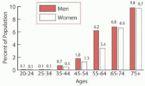
FIGURE 35.1. Prevalence of congestive heart failure by age and sex. Zoghbi WA, Enriquez-Sarano M, Foster E. et al. Recommendations for evaluation of the severity of native valvular regurgitation with two-dimensional and Doppler echocardiography. J Am Soc Echocardiogr 2003;16(7):777-802.

Strategies for treating end-stage CHF aim to improve quality of life, limit disease progression, and prolong life. Medical therapies, such as angiotensin-converting enzyme inhibitors (ACEI), β blockers, diuretics, inotropic agents, and antiarrhythmics, represent the usual standard of care for CHF management. However, even multidrug regimens may not prevent progression toward end-stage CHF; when this occurs, surgery is the only effective intervention.

Cardiac transplantation is the “gold-standard” surgical treatment for end-stage CHF. While the one-year survival rate after transplantation is 85%, patients in New York Heart Association (NYHA) class IV have a one-year mortality rate of 40%-50% (5,6). Additionally, the small number of donors limits the number of CHF patients who can benefit from cardiac transplantation. Over the past decade, the number of transplant recipients older than 65 years has quadrupled, but the number of donors has remained the same and transplantation waiting times have increased steadily. As a result, even though almost 16,000 people per year could benefit from cardiac transplantation, only 2,400 heart transplantations are actually performed.7 The mismatch between the growing number of cardiac transplant candidates and the limited number of donors has led to increasing use of alternative advanced surgical therapies intended to unload the heart and allow myocardial reverse-remodeling and recovery. Included among these advanced procedures are mechanical ventricular assist device (VAD) implantation, ventricular remodeling procedures in conjunction with revascularization and mitral valvuloplasty (i.e., endoventricular circular patch plasty (ECPP), partial ventriculectomy), newer procedures intended to reduce ventricular dilatation (i.e., external or internal splinting), and total artificial heart (TAH) implantation.


CRITICAL ISSUES ADDRESSED BY INTRAOPERATIVE ECHOCARDIOGRAPHY IN PATIENTS UNDERGOING PROCEDURES FOR CONGESTIVE HEART FAILURE

Intraoperative echocardiography (IOE) (consisting of both transesophageal echocardiography (TEE) and epicardial echocardiography) can be used to identify the important aspects of a particular case of end-stage CHF and promote successful surgical intervention (Table 35.2).


Determine the Etiology and Mechanism of CHF

Advanced CHF is caused by a variety of diseases that affect the myocardium. Based on their functional and morphologic features, cardiomyopathies (CMP) can be classified as dilated, hypertrophic, or restrictive. Dilated CMP is the result of viral, bacterial, or parasitic disease of toxic insult. It can also occur in association with pregnancy. Long-term severe myocardial ischemia and chronic regurgitant valvular disease, such as mitral or aortic insufficiency, could also lead to dilated CMP (Figs. 35.2 and 35.3). Hypertrophic CMP is either genetically inherited or is the result of long-standing hypertension or valvular disease, such as aortic stenosis. Infiltrative processes (amyloidosis, hemochromatosis) or inflammatory disease (sarcoidosis) are the most common causes of restrictive CMP. IOE is an important diagnostic tool due to its unique ability to evaluate cardiac morphology and function and to differentiate among various types of CMP (Tables 35.3 and 35.4) (8).


Assessment of Left and Right Ventricular Function

Assessment of left ventricular (LV) and right ventricular (RV) function is critical to the proper evaluation and perioperative management of patients with end-stage CHF. Using IOE can provide excellent qualitative and quantitative information on RV and LV function.

Dilated CMP is characterized by dilation of both ventricular and atrial chambers associated with systolic heart failure (9). Both motion mode (M-mode) and two-dimensional (2-D) echocardiography can be used for the evaluation of LV systolic function. The high time resolution of M-mode echocardiography enables it to accurately measure ventricular internal dimension and wall thickness. Additionally, placing the M-mode beam at the mitral chordal level to obtain a transgastric longitudinal view of the LV makes it possible to compute LV fractional shortening,
a rough measurement of LV systolic function with a normal range of 25% to 45%, using the following formula:


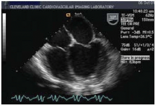
FIGURE 35.2. Dilated cardiomyopathy: midesophageal four-chamber view


TABLE 35.2. Application of Intraoperative Echocardiography During Surgical Procedures for Congestive Heart Failure


Surgical Procedure


Critical IOE Issues


Pre-CPB


Post-CPB


LVAD Implantation


RV function


Sequelae of CHF MR, pulmonary HTN Diastolic dysfunction Mural thrombi (LA, LV)


Potential R → L shunt


Potential Ao → LV circuit


Cannulation-perfusion


LVAD cannula inflow/outflow Position and function


LVAD function LV volume Valve competence


Complications of CPB


Global RV Fx, RV FAC


MR (grade, mechanism)


LAA or LV mural thrombi


PW of MV Inflow pulm vein contrast


CFD, contrast with valsalva


CFD of AV


Global RV Fx, RV FAC


PW of MV and pulmonary vein


CFD of AV


TEE and EPE


Apical inflow position CW, CFD


Aortic outflow Air embolism


Hypovolemia, flow < 1.5 m/sec


Regurgitant LVAD flow


2-D Asc and descending aorta


BI-VAD, ECMO


RV Function


Sequelae of CHF MR, pulmonary HTN Diastolic dysfunction Mural thrombi (LA, LV)


Potential R → L shunt


Potential Ao → LV circuit


Cannulation-perfusion


LVAD Cannula inflow/outflow Position and function


LVAD function LV volume Valve competence


Complications of CPB cannula positioning LA, PA, LV, Ao


LA thrombus formation


Global RV Fx, RV FAC


PW of MV and pulmonary vein


CFD of AV


TEE and EPE


Apical inflow position CW, CFD


Aortic outflow Air embolism


Hypovolemia, flow < 1.5 m/sec


Regurgitant LVAD flow


2-D Asc and descending aorta


Dor Procedure


Ventricular Reconstruction Remodeling LV CABG MV Repair


RV function


LV function and scar


Sequelae of CHF MR, pulmonary HTN Diastolic dysfunction Mural thrombi


Potential R → L shunt


Embolic source (LA, LV)


Cannulation-perfusion strategy


Myocardial viability


MR severity and mechanism


Global RV Fx, RV FAC, TR


RWMA, scar


MR (severity and mechanism)


PWD MW and pulmonary vein contrast


CFD, contrast and valsalva


EPE


Resting RWMA


Low-dose dobutamine stress


Global RV Fx, RV FAC


PW of MV and pulmonary vein


CFD of AV


TEE and EPE


Apical inflow position CW, CFD


Aortic outflow Air embolism


Hypovolemia, flow < 1.5 m/sec


Regurgitant LVAD flow


2-D Asc & descending aorta


Revascularization


Myocardial viability


Resting RWM


Low-dose dobutamine stress


Mitral valve function


Assess for new RWMA


Comprehensive IOE focusing on complications of cannulation, perfusion, myocardial protection, and valve function


MV Repair


Mitral regurgitation


MR severity and mechanism


Pathoanatomy, annulus size


Success of repair


Complications


External and Internal Splinting


Size LV, Systolic LV / RV Fx, MR, diastolic Fx


Position of mechanical splints


As above, LV dimensions, LV & RV dimensions, Ej Fx MR mechanism/severity, mitral inflow, pulmonary vein CW


Diastolic function Filling RV function


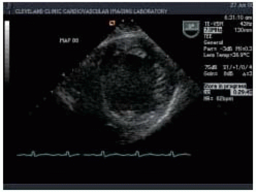
FIGURE 35.3. Dilated cardiomyopathy: transgastric midpapillary short-axis view

Fractional shortening (%) = (LVIDd − LVIDs)/LVIDd × 100%

(LVIDd = left ventricular internal diameter at enddiastole; LVIDs = left ventricular internal diameter at end-systole).

Two-dimensional echocardiography provides both qualitative and quantitative evaluation of systolic ventricular function. Midesophageal four-chamber (ME 4-chamber) and two-chamber (ME 2-chamber) views and the midesophageal long-axis (ME LAX) view, transgastric short-axis (TG SAX) views (basal, midpapillary, and apical), and the transgastric long-axis view (TG LAX) allow assessment of both global and regional ventricular function. Two-dimensional quantitative measures of LV systolic function include ventricular dimensions, volume, stroke volume (SV), cardiac output (CO), ejection fraction (EF), and regional wall motion abnormalities (Figs. 35.4 and 35.5). Left ventricular ejection fraction (LVEF) can be calculated from LV end-systolic volume (LVESV) and end-diastolic volume (LVEDV) as the ratio (LVEDV-LVESV)/LVEDV. Cardiac output can be calculated using the area-length formula across a cardiac valve (most commonly the aortic valve):


TABLE 35.3. Typical Features of the Three Physiologic Types of Cardiomyopathy


Dilated


Hypertrophic


Restrictive


LV systolic function


LV diastolic function


LV hypertrophy


Moderately to severely ↓


May be abnormal


↑ LV mass due to left


ventricular dilation with


normal wall thickness


Normal


Abnormal


Asymmetric LV hypertrophy


Normal


Abnormal


Concentric LV hypertrophy


Chamber dilation


All four chambers


Left and right atrial dilation if mitral regurgitation is present


Left and right atrial dilation


Outflow tract obstruction


Absent


Dynamic LV outflow tract obstruction may be present


Absent


Left ventricular end-diastolic pressure


Elevated


Elevated


Elevated


Pulmonary artery pressures


Elevated


Elevated


Elevated


LV = Left ventricular; ↓ = decreased; ↑ = increased.


From Otto CM. The cardiomyopathies, hypertensive heart disease, post-cardiac-transplant patient and pulmonary heart disease. Textbook of Clinical Echocardiography, Philadelphia: WB Saunders 2000;183-212.


CO = 0.785 × LVOT D2 × LVOTVTI × HR

(LVOT = left ventricular outflow tract; D = LVOT diameter; LVOTVTI = velocity time integral, measured with Doppler spectrum across the aortic valve; HR = heart rate). IOE determination of CO is helpful, even when a pulmonary artery catheter is used, because some patients with dilated CMP present with some degree of tricuspid valve regurgitation, which renders the thermodilution technique inaccurate.

In addition, patients with end-stage CHF often present with coexisting pathology such as acquired left ventricle aneurysm (LVA). LVA is the final expression of transmural infarct expansion and is often accompanied by anterior infarction in the area of the distribution of the left anterior descending coronary artery (Fig. 35.6). Thrombi are frequently detected within aneurysms and are often associated with systemic embolization (Fig. 35.7). Therefore,
careful echocardiographic assessment is mandatory if cardiopulmonary bypass is to be successful.


TABLE 35.4. Echocardiographic Approach to the Patient with a Suspected Cardiomyopathy


Qualitative


Quantitative


2-D/M-mode imaging


Chamber dimensions


Degree and pattern of LV hypertrophy


Evidence for dynamic outflow tract obstruction


SAM of mitral valve


Aortic valve midsystolic closure


LV-EDV, LV-ESV


LV mass


LV systolic function


Doppler echo


Visual estimate of EF


Associated valvular regurgitation


Pattern of LV diastolic filling


Pattern of LA filling (pulmonary venous inflow)


Velocity curve of dynamic outflow tract obstruction


Localization of the level of obstruction


Apical biplane EF


Maximum and mean ΔP


PA pressures


2-D, Two-dimensional; LA, left atrial; LV, left ventricular; EDV, end-diastolic volume;


ESV, end-systolic volume; SAM, systolic anterior motion; ΔP, pressure gradient; PA, pulmonary artery; EF, ejection fraction.


Otto CM: The cardiomyopathies, hypertensive heart disease, post-cardiac-transplant patient and pulmonary heart disease. Textbook of Clinical Echocardiography, W.B. Saunders 2000,183-212.


Evaluation of RV systolic function is a critical part of the intraoperative management of patients with end-stage CHF. Preexisting pathologic conditions in the RV, such as ischemia, infarction, CMP, and pulmonary hypertension (PHTN) are major risk factors for RV failure. Sometimes, the extent of RV dysfunction becomes apparent only when a sudden increase in venous return challenges an already impaired RV. With dilated CMP, there is an increase in systolic ventricular interaction, making RV systolic performance more dependent on the LV to generate pressure (10). Under these circumstances, sudden LV unloading may depress RV function further.


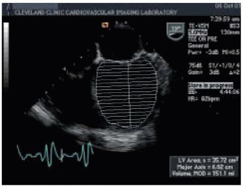
FIGURE 35.4. Dilated cardiomyopathy, left ventricle dimensions and volume: midesophageal four-chamber view

Two-dimensional/M-mode echocardiography can provide several quantitative and qualitative measurements of RV systolic function. Wall thicknesses over 0.5 cm as measured by M-mode echocardiography are considered abnormal and suggest elevated pulmonary artery pressure, pulmonary valve stenosis, or infitrative CMP. The standard 2-D transesophageal echocardiographic (TEE) views used to evaluate RV function are the ME 4-chamber view, the ME RV inflow-outflow view, the transgastric mid-short-axis (TG mid-SAX) view, and the transgastric right ventricular inflow (TG RV inflow) view. Quantitative assessment of RV function with automated border detection in the midesophageal four-chamber view has been used to calculate RV fractional area change (RVFAC) (11):


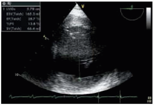
FIGURE 35.5. Dilated cardiomyopathy, left ventricle dimension and volume: transgastric short-axis midpapillary view


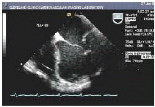
FIGURE 35.6. Left ventricular aneurysm: midesophageal four-chamber view

RVFAC = (end-diastolic area) − (end-systolic area/end-diastolic area) × 100%

Signs of RV dysfunction include hypokinesis or akinesis of the RV free wall, and RV dilation caused by volume or pressure overload. Normally, the RV end-diastolic cross-sectional area is less than 60% of the LV end-diastolic cross-sectional area. With dilatation, however, the RV changes its shape from triangular to round, with concomitant enlargement of the right ventricular outflow tract (RVOT) and flattening of the bulge in the interventricular septum from right to left can be seen in the ME 4-chamber view and the ME RV inflow-outflow view (Figs. 35.8 and 35.9). With RV pressure overload, a maximal leftward septal shift is noted at end-systole, whereas RV volume overload is associated with a maximum reversed septal curvature in mid-diastole. As RV dilation becomes moderate or severe, the RV replaces the LV in forming the cardiac apex in the ME 4-chamber view, and the end-diastolic cross-sectional area of the RV may equal or exceed that of the LV. Tricuspid annular plane systolic excursion (TAPSE) may decrease to less than 20 mm, accompanied by impaired RV systolic function (12). At this stage, pulsed wave Doppler (PWD) evaluation of blood flow in the hepatic vein may reveal attenuation of the systolic inflow wave.


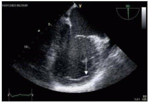
FIGURE 35.7. Ventricular thrombus: midesophageal two-chamber view


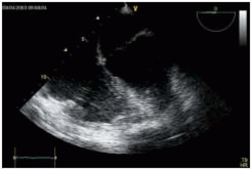
FIGURE 35.8. Biventricular failure: midesophageal four-chamber view

Nonetheless, pulmonary artery (PA) pressure estimates remain one of the most important quantitative measures of RV systolic function. As RV dysfunction progresses, dilation of tricuspid valve (TV) annulus occurs, causing varying degrees of tricuspid regurgitation (Fig. 35.10). Measuring the velocity and pressure gradient of the tricuspid regurgitant jet (Fig. 35.11) reveals the gradient between RV and right atrial (RA) pressure and, when added to an estimate of RA pressure (transduced from the central venous line), allows calculation of RV systolic pressure
(Table 35.5). Right ventricular SV and CO can be measured directly by imaging the RVOT tract and PA in the upper esophageal aortic arch short-axis (UE aortic arch SAX) view (Fig. 35.12). By measuring the diameter (D) of PA annulus and using PWD mode to determine velocity time integral (VTI) across pulmonic valve, right ventricular SV, and CO can be calculated using the formulas SV = PA D2 × 0.785 × PAVTI and CO = SV × HR.


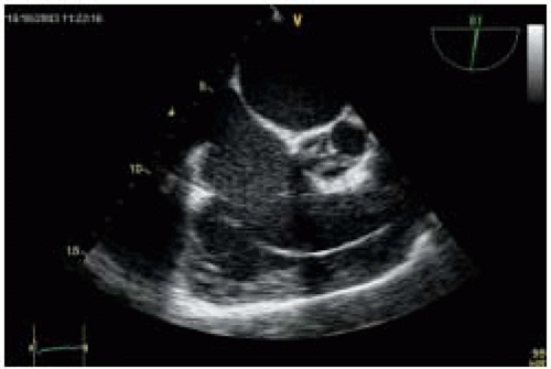
FIGURE 35.9. Right ventricular failure: midesophageal right ventricular inflow-outflow view


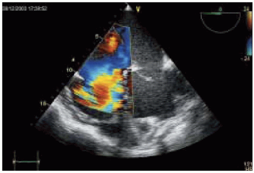
FIGURE 35.10. Severe tricuspid regurgitation: midesophageal four-chamber view


TABLE 35.5. Estimation of Intracardiac Pressures


Pressure Estimated


Required Measurement


Formula


Normal Valves (mm Hg)


Estimated CVP


Respiratory IVC collapse


> 40% = 5 mm Hg < 40%, (nl RV) = 10 mm Hg 0% & RV Dysfx = 15 mm Hg


5-10


RV Systolic(RVSP)


Peak velocityTR CVP or measured


RVSP = 4(VTR)2 + CVP (No PS)


16 – 30


RV Systolic (with VSD)


Systemic systolic BP Peak V LV-RV


RVSP = SBP – 4(V LV-RV)2 (No LVOT Obstruction)


with VSD usually > 50


PA Systolic PASP


Peak velocityTR CVP* or measured


PASP = 4(VTR)2 + CVP No PS)


16 – 30


PA Diastolic (PAD)


End diastolic VelocityPR CVP* or measured


PAEDP = 4(VPR ED)2 + CVP


0 – 8


PA Mean (PAM)


Acceleration time (AT) to peak VPA


PAM = (-0.45) AT + 79


10 – 16


RV dP/dt


TR spectral envelope


T TR(2m/sec) – T TR(1 m/sec)


RV dP = 4V2 TR(2m/sec) – 4V2TR(2m/sec)


RV dP/dt = dP / T TR(2m/sec) – T TR(1 m/sec)


> 150 mm Hg/msec


LA Systolic (LASP)


Peak VMR Systolic BP (SBP)


LASP = SBP – 4(VMR)2 (No LVOT Gradient)


100 – 140


LA (PFO)


VelocityPFO CVP* or measured


LAP = 4(VPFO)2 + CVP


3 – 15


LV Diastolic (LVEDP)


End diastolic


VelocityAR


Diastolic BP (DBP)


LVEDP = DBP – 4(VAR)2


3 – 12


LV dP/dt


MR spectral envelope T MR(2m/sec) – T MR (1 m/sec)


LV dP = 4V2 TR(2m/sec) -4V2TR(2m/sec) LV dP/dt = dP / T MR (2m/sec) – T MR (1 m/sec)


> 800 mm HG/msec


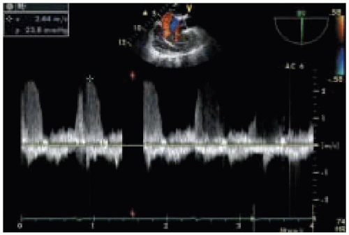
FIGURE 35.11. Tricuspid regurgitation, velocity, and pressure gradient

Growing evidence suggests that both systolic and diastolic dysfunction play important roles in the clinical presentation and prognosis of patients with end-stage CHF. Patients with hypertrophic, infiltrative, or primary restrictive
CMP may report symptoms of heart failure despite normal EF, a condition known as diastolic heart failure (13,14). In addition, in patients with preexisting systolic dysfunction, abnormalities in diastolic dysfunction may be significantly related to the severity of cardiac symptoms and prognosis in patients with CHF (15). Diastole is the interval between aortic valve closure and mitral valve closure and can be divided into four phases:


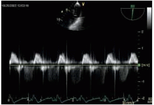
FIGURE 35.12. Pulsed wave Doppler of pulmonary artery

1. Isovolumic relaxation

2. Early rapid diastolic filling

3. Diastasis

4. Late diastolic filling caused by atrial contraction (Fig. 35.13).

Parameters of diastolic function include ventricular relaxation, myocardial compliance, and chamber compliance. Ventricular relaxation is measured by isovolumic relaxation time (IVRT), the rate of pressure decline (dP/dT), and time constant of relaxation (τ). Myocardial compliance is estimated by the ratio of change in volume to change in pressure (dV/dP). Chamber compliance is assessed by measuring early diastolic LV filling (E wave), deceleration time (DT), late diastolic LV filling (AM wave), the AP wave (reversed) of LA contraction, the s wave of the systolic LA filling phase, and the d wave of the LA diastolic filling phase.

The earliest stage of abnormal diastolic filling is impaired relaxation with inverse E/A ratio as the major Doppler abnormality (Table 35.6). With progression of the disease to moderate diastolic dysfunction, pseudonormalization of diastolic filling flow occurs because of impaired myocardial relaxation balanced by elevation of mean LA pressures (Table 35.6). The diagnosis is confirmed by abnormal PV flow or response to Valsalva maneuver. The restrictive filling pattern is the most advanced form of diastolic dysfunction that can be associated with either normal or abnormal systolic function. It may accompany advanced infiltrative CMP, such as amyloidosis, advanced hypertensive disease, or dilated CMP. The hallmark of the disease is elevated LA pressures with increased LV stiffness that causes a large E wave, short DT, a small AM wave, and a very small s/d ratio on PV PWD trace (Figs. 35.14 and 35.15) (Table 35.7) (16).


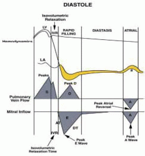
FIGURE 35.13. The relationship among left ventricular, left atrial, and aortic pressures


Assessment of Coexisting or Secondary Pathology

Mitral regurgitation (MR) is commonly encountered in CHF as a consequence of dilated cardiomyopathy (17,18,19). Mitral annular dilation caused by LV dilation and papillary muscle dysfunction are the most common mechanisms behind the development of regurgitation in dilated cardiomyopathy. Abnormal papillary muscle alignment and abnormal leaflet apposition during systole are caused by changes in the shape of the LV chamber during both the systolic and the diastolic periods (20). Color flow Doppler (CFD) imaging has been used to quantitate the severity of MR, whereas PWD is used to measure regurgitant stroke volume and regurgitant fraction (21,22). Tricuspid regurgitation (TR) can be encountered either in isolation or in conjunction with MR. The regurgitant tricuspid jet is usually directed toward the interatrial septum in patients with dilated CMP. RV dysfunction, PHTN, patent foramen ovale (PFO), and aortic regurgitation (AR) are other pathophysiologies that may accompany end-stage CHF.


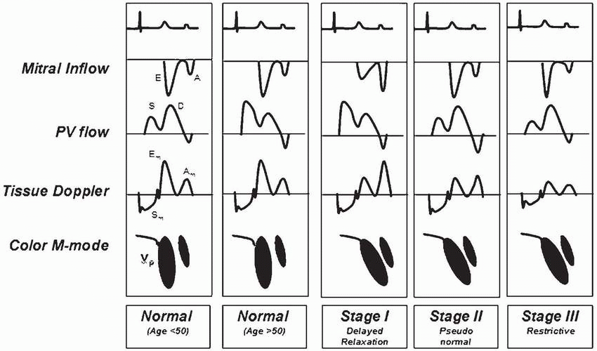
TABLE 35.6. Patterns of Diastolic Dysfunction


image


Standard Intraoperative Echocardiographic Examination

IOE is a key monitoring and diagnostic modality in patients undergoing surgery for heart failure. Standard IOE evaluation usually begins with the ME 4-chamber view. This view provides information about the size of the left and right cardiac chambers; the thickness of ventricular walls and the basal, mid, and apical segments of LV lateral and septal walls; the apical and basal portion of RV free wall; and MV and TV function. PWD assessment of the left upper and lower pulmonary veins (LUPV, LLPV), the right upper pulmonary vein (RUPV), and MV diastolic flow can be used to estimate the severity of MR and the extent of diastolic impairment. Next, the multiplane angle is rotated 30° into the ME AV SAX view, which allows examination of morphology and function of the AV cusps. The ME RV inflow-outflow view is obtained at 60° and provides information about RV diaphragmatic free wall motion, RVOT size, and pulmonary valve (PV) and TV function. The ME mitral commissural view provides information about the A2 scallop of the anterior mitral valve leaflet (AMVL) and the P1 and P2 scallops of the posterior mitral valve leaflet (PMVL) and permits assessment of the severity and direction of the MV regurgitant
jet, if present. The ME 2-chamber view at 90° allows evaluation of the basal, mid, and apical segments of the anterior and inferior LV walls as well as examination of the left atrial appendage (LAA) for mural thrombi. The ME LAX view at 120 shows both basal and midanteroseptal segments, basal and midposterior segments, the left ventricular outflow tract (LVOT), and the aortic valve. The ME bicaval view is the image of choice for evaluating PFO by CFD and contrast study. This view provides information about the size of and blood flow through the RA, the left atrium (LA), and the right lower pulmonary vein (RLPV) using PW Doppler. TG views are obtained by advancing the probe into the stomach and flexing the tip anteriorly. TG basal and mid-SAX views are the views of choice for the examination of LV regional wall motion abnormalities. In addition, the TG basal SAX view provides a short-axis view of the MV that allows further location of the regurgitant jets using CFD. Rotating the multiplane angle 90° to the TG 2-chamber view allows further assessment of the LV anterior and posterior walls, the MV, and the LAA. The TG LAX view at 120° provides a longitudinal view of the AV and allows measurement of pressure gradients across the AV using continuous wave Doppler (CWD) and PWD. Turning the probe to the right and to 120° to the TG RV inflow view allows evaluation of the RV diaphragmatic and free walls, TV function, and RA dimensions. The deep TG LAX view provides a longitudinal view of the LV and AV and allows measurement of AV gradients and calculation of the AV area using CWD and PWD. Atherosclerotic disease in the descending aorta can be detected with the probe in a gastric position and the tip rotated posteriorly into the left descending aortic short-axis (descending aortic SAX) view. A longitudinal view of the descending aorta can be obtained at 90 in the descending aortic long axis (descending aortic LAX) view. The ME ascending aortic SAX/LAX views and upper esophageal (UE) aortic arch LAX/SAX views are used to examine the ascending aorta and should be used to guide cannulation and cross-clamping of the aorta. Doppler examination of the pulmonary valve is best achieved using the UE aortic arch SAX view (23).


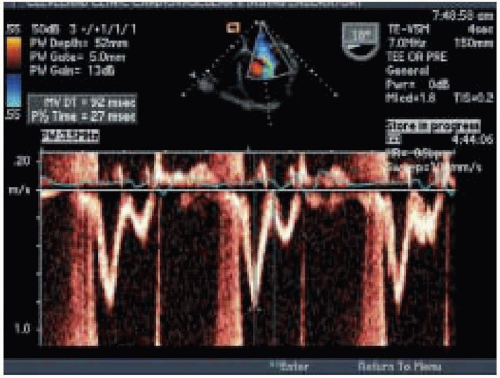
FIGURE 35.14. Pulsed wave Doppler: transmitral diastolic flow


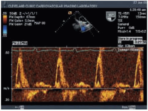
FIGURE 35.15. Left upper pulmonary vein pulse wave Doppler: restrictive pattern


TABLE 35.7. Distinguishing the Patterns of Diastolic Dysfunction


Parameter


Normal
Filling


Impaired
Filling
Stage 1


Pseudonormal
Filling
Stage 2


Restrictive
Filling
Stages 3 and 4


E Wave


DT


160 msec-240 msec


> 240 msec


160 msec-200 msec


< 160 msec


IVRT


70 msec-90 msec


> 90 msec


< 90 msec


< 70 msec


E : A


1-2


<1


1-1.5


>1.5


AM : AP Duration


AM ≥ AP


AM > AP


AM < AP


AM << AP


PVS : PVD


PVS > PVD


PVS : >> PVD


PVS < PVD


PVS << PVD


VE’ MA : VA’ MA


VE’ MA > VA’ MA


VE’ MA < VA’ MA


VE’ MA < VA’ MA


VE’ MA < VA’ MA


Valsalva


Decreased


Decreased


Reversal


Decreased


E : A


<1


Volume Loading


AM < AP


AM << AP


AM <<< AP


AM : AP Duration


Surgical Procedure-Related Issues

Surgical approaches to end-stage CHF are rapidly becoming more numerous and sophisticated. Substantial advances have been made in myocardial revascularization, mitral valvuloplasty, ventricular remodeling (i.e., ECPP and LV partial resection), ventricular constraint techniques (i.e., Acorn CorCap external splinting, Myosplint internal splinting), mechanical ventricular support, TAH, and heart transplantation. During the period before cardiopulmonary bypass (CBP) is initiated, IOE focuses on the evaluation of

1. RV function, severity of TR, and the potential need for RV mechanical support

2. Baseline LV function

3. Potential sequelae of end-stage-dilated cardiomyopathy, such as LVA, LA mural thrombi, MR, and diastolic dysfunction

4. Potential right-to-left shunting through PFO, which may contribute to hypoxemia

5. The presence of AR or mitral stenosis (MS)

6. The presence of ascending aortic atheroma, which necessitates changing the cannulation strategy and myocardial protection.

This information guides the development of CPB strategy. At the time of separation from CPB, IOE is useful for evaluating the adequacy of deairing of the cardiac chambers. It is also important at that time to establish a comprehensive baseline study for future comparison and to assess LV and RV filling patterns, native valve function, the quality of the valve repair, and the presence of any intracardiac shunts. Given the importance of RV function in the outcome of the procedure, an objective qualitative assessment of RV systolic and diastolic function by RVFAC,
RV end-diastolic volume (RVEDV), RV dP/dT, TV annulus diameter, and severity of TR is important. These parameters will allow comparison between baseline and postoperative RV performance and dictate postoperative management in the event of acute deterioration of RV function, which is often seen when respiratory insufficiency occurs.


Complications of Surgical Procedures for CHF


Right Ventricular Dysfunction

The success of surgical procedures for end-stage CHF depends on postoperative RV performance. Unfortunately, right-sided circulatory failure is a common postoperative complication in patients undergoing surgical procedures for CHF. Patients with end-stage CHF often develop substantial passive pulmonary hypertension secondary to elevated LA and pulmonary venous pressures, a condition that is reversible with LV unloading. When pulmonary disease is also present, irreversible elevated pulmonary vascular resistance (PVR) complicates the clinical picture. RV function is significantly afterload dependent, and the presence of pulmonary congestion in association with high PVR can have a tremendous impact on RV systolic and diastolic performance. RV failure causes dilation, ischemia, and decreased RV contractility. It is associated with decreased pulmonary blood flow and a leftward septal shift that subsequently lowers LV filling pressure and reduces systemic CO. Treatment of RV is difficult (Table 35.8).

Only gold members can continue reading. Log In or Register to continue

Stay updated, free articles. Join our Telegram channel

Jul 15, 2016 | Posted by in CARDIOLOGY | Comments Off on Assessment in Surgical Procedures for Congestive Heart Failure

Full access? Get Clinical Tree

Get Clinical Tree app for offline access