Assessment in Higher Risk Myocardial Revascularization and Complications of Ischemic Heart Disease


Assessment in Higher Risk Myocardial Revascularization and Complications of Ischemic Heart Disease


Robert M. Savage

Gonzalo Gonzalez-Stawinski

Jacek Cywinski

David Vener

Bruce W. Lytle


HISTORICAL PERSPECTIVES

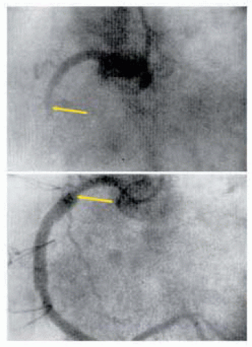
The first association between coronary artery disease and myocardial dysfunction was suggested in 1779 by Caleb Hillier Parry who described his autopsy findings of hardened “ossified” blockages in the coronary arteries and death associated with “syncope anginos” (1). It was three years later that William Heberden provided his classic description of the syndrome of pectoralis dolor as “a disagreeable sensation in the breast” associated with exertion (2). In 1856, Rudolf Virchow described the evolution of fibrous thickening in the arterial wall into arterial atheroma because of a reactive inflammatory process resulting in fibrotic proliferation cells (3). Since these early characterizations of coronary artery disease, remarkable advances have been made in our understanding of this disease process and its management. In 1910, Alexis Carrell performed the first aortocoronary bypass surgery in a dog, using a preserved carotid artery between the ascending aorta and left anterior descending coronary artery (4). However, it was not until early 1958 that Longmire was credited with performing the first internal mammary to coronary artery anastomosis following a right coronary endarterectomy, which had “disintegrated” (5). As seen in Tables 24.1 and 24.2, the management of coronary artery disease has been driven by numerous factors and spans more than two centuries from initial scientific observations to the advanced direct surgical and percutaneous approaches to myocardial revascularization. While there may be a number of surgical teams credited with performing the first coronary artery bypass surgery, it was René Favaloro’s report, in 1968, of 171 patients undergoing direct surgical myocardial revascularization that ushered in the modern era of coronary bypass surgery (Fig. 24.1) (6). Since that time, there have been number of milestones in the evolution of myocardial revascularization.

1. The introduction of myocardial protection strategies

2. Demonstration of long-term advantages of IMA conduits

3. Advances in cardiovascular anesthetic management

4. Introduction of percutaneous approaches to revasculrization

5. Introduction of intraoperative echocardiography into patient management

6. Improved management of the complications of ischemic heart disease

7. Identification of the variables improving repeat coronary bypass surgery

8. Development of ventricular support devices

9. Development of ventricular reconstruction surgery


INDICATIONS FOR MYOCARDIAL REVASCULARIZATION

The American College of Cardiology recently revised their Guidelines for Coronary Artery Bypass Graft Surgery (CABG) (Table 24.3) (7). A number of factors created an evolution in the current indications for myocardial revascularization by traditional surgical CABG or percutaneous coronary intervention (PCI). The factors are related to patient selection, differences in institutional CABG and PCI capabilities, temporal differences in recurrence for CABG and PCI, accelerated pace of innovations affecting the scientific study relevancy, and the inherent difficulties in comparing CABG and PCI due to the inability to account for all of the definable and less definable outcome risks in randomized studies (7,8). While the pace may slow, physicians involved in the care of patients with coronary artery disease will need to stay current with the scientific literature and ongoing innovations in revascularization.


DEFINITION OF HIGHER RISKS IN PATIENTS UNDERGOING CORONARY ARTERY BYPASS GRAFT SURGERY

To assist patients in making an informed decision and enable the surgical team to develop optimal perioperative strategies, it is important to identify factors that may affect a patient’s perioperative outcome. To identify risks of morbidity and mortality, patient and disease-related features from patient’s history, physical examination, or diagnostic evaluation are subjected to statistical analysis to determine the degree of correlation with adverse clinical events. Such risks are related to the severity and extent of ischemic heart disease or other comorbid chronic disease processes. While many factors are associated with higher morbidity and mortality, it is typically only those commonly occurring that permit a statistical correlation with outcomes. From these studies, outcome prediction
may be based on cumulative risks (7). While many of these risks are easily defined (Table 24.3), others are more difficult to define objectively. Yet, many of these issues have a significant impact on the patient’s outcome. Included in these more difficult to define risks are:


TABLE 24.1. History of Coronary Artery Disease and Its Management


Year


Investigator


Milestone


1770-1935


Observations Considerations for Treatment


1779


Caleb Hillier Parry


Discovered relation between angina and coronary ossification


1856


Rudolf Virchow


Described inflammatory process of atheroma development


1856


William Heberden


Characterized classic angina


1880


Langer


Described coronary collateral communications


1899


Francois-Franck


Described sympathetic innervation


1902


Kocher


Absence of angina in thyroidectomy patient


1910


Alexis Carrell


Experimental aortocoronary bypass with preserved carotid artery


1916


Jonnesco


Performed first cardiac sympathetectomy


1926


Boas


Subtotal thyroidectomy for treatment of angina


1929


Richardson and White


Series of patients undergoing ganglionic sympathetectomy


1930


Sussman


Performed cardiac irradiation for sympathetic denervation


1930s


Carrell and Lindberg


Developed primitive heart lung machine


1935-1953


Indirect myocardial revascularization


1930


Claude S. Beck


Performed epicardial abrasion to increase collateral flow


1934


Robertson


Ligated coronary sinus to redirect coronary flow


1937


O’Shaughnessy


Used omental flap to epicardium for revascularization


1937


John Gibbon, Jr.


Bypassed a dog’s heart during pulmonary artery occlusion


1938


Griffith and Bates


Direct implantation of blood vessels into myocardium


1946


Arthur Vineberg


Reported internal mammary implants directly into myocardium


1946


Beck


Arterialized coronary sinus


1951


Gordon Murray


Direct arterial repair and venous interposition homografts


1953


William Mustard


Carotid to coronary bypass


1953


John Gibbon


First effective heart-lung bypass machinery


1954-1966


Early direct coronary artery bypass


1954


Murray


First successful bypass on beating heart of a dog


1955


Melrose


Elective potassium-induced arrest


1957


F. Mason Sones


First cineangiogram of coronary artery


1958


William Longmire


Grafted IMA to coronary vessel


1958


Senning


Coronary endartectomy with plaque excision and graft


1960 (reported 1964)


Robert Goetz


IMA to right coronary anastomosis


1962 (reported 1974)


David Sabiston


First saphenous vein CABG


1964


Vasilii Kolesov


Performed internal mammary to LAD graft without CPB


1964 (reported 1973)


Garrett and DeBakey


First successful saphenous vein bypass graft


1966


Bailey


Gastroepiploic artery implantation into myocardium


1967-present


Modern era of revascularization


1967


René Favaloro


First series of free SVG and end-to-side anastomosis


1968


Dudley Johnson


Other saphenous vein grafting series


1970


René Favaloro


Double IMA grafts alone or in combination with SVG


1968


René Favaloro


Aortocoronary bypass for unstable angina and AMI


1968


René Favaloro


Combined CABG and valve replacement or aneurysmectomy


1973


Alan Carpentier


Free radial artery grafts


1974


Gerald Buckberg


Myocardial protection strategies using cardioplegia


1976-1986


Floyd Loop


Reported survival benefit IMA-LAD graft


Mueller RL, Rosengart TK, Isom W. The history of surgery for ischemic heart disease. Ann Thorac Surg 1997;63:869-78.


FIGURE 24.1. René Favaloro and Mason Sones of the Cleveland Clinic united as a team in demonstrating the feasibility of safely performing saphenous vein interposition and aortocoronary bypass grafts. In May of 1967, this angiogram demonstrated the intersegmental graft with end-to-end anastomoses. In December of 1968, Favaloro, Sones, and Effler summarized the advances in the first large series of 171 patients. Favaloro, RG. Landmarks in the development of coronary bypass surgery. Circulation. 1998;98(5):466-78.

1. Availability of suitable bypass conduits

2. Diffuseness of distal coronary atherosclerosis (reducing distal coronary blood flow)

3. Presense of noncardiac atherosclerosis

4. Uncommon combinations of patient comorbidities (9,10)


Risk Factors and Mortality

There have been a number of studies evaluating the clinical variables associated with perioperative death (11,12,13). They are difficult to compare due to differences in the definitions of clinical or disease variables, clinical endpoints, institutional differences in clinical practices, and clinical outcomes. Jones et al. combined the data from seven large studies to evaluate recurring factors that contribute to in-hospital mortality following CABG (14a). This resulted in a clinical study population of more than 172,000 patients, enabling the identification of two levels of variables. Those variables having the strongest correlation with outcome included:


TABLE 24.2. Challenges with Guideline Indications for Myocardial Revascularization Using CABG and PCI


1.


Rapid pace of technology


2.


Time between scientific investigation and technology


3.


Time difference in disease recurrence for CABG and PCI


4.


Comparability of CABG-PCI study groups (selection bias, nonrandomized patient, and disease risk factors)


5.


Randomized trials for degree of risk


6.


Initial therapy studies include crossover success


7.


Differences in definitions of re-stenosis in CABG and PCI


8.


Secondary prevention in studies differs


9.


High volume center studies not as relevant to low volume practices


10.


Unique capabilities of individual centers in CABG and PCI


11.


Difficulties of informed consent


1. Patient’s age

2. Gender

3. Previous cardiac surgery

4. Operation urgency

5. Ventricular ejection fraction

6. Characterization of coronary anatomy (left main > 50% stenosis, number of vessels with > 70% stenosis)

Age, urgency of procedure, and reoperation were the variables most strongly correlated with mortality. There were other variables identified that influenced mortality but they were not as strongly correlated (Table 24.4).

Trials that were initiated in the first decade of coronary bypass surgery demonstrated that patients with left main or triple vessel disease and abnormal LV function had an improved long-term survival when coupled with an aggressive strategy of complete revascularization (14b,14c). Since then, there have been remarkable improvements in the management of this unique group of higher risk patients with the advent of myocardial protection strategies, integrated perioperative care, secondary prevention, use of arterial grafts, and the increased collective experience of the surgical community. This has led to a growing consensus, as outlined by the recent ACC Guidelines for Coronary Artery Bypass Surgery, that left main equivalent or triple vessel disease combined with abnormal LV function are indications for surgical revascularization (Table 24.2) (7). Despite recent improvements in the medical management of this group of patients, the yearly mortality remains at 12%, indicating
that surgical management of this high-risk group is warranted (14d). This strategy is further supported by an understanding that:


TABLE 24.3 Indications for Coronary Artery Bypass Surgery


Asymptomatic or Mild Angina


Class I


1.


Left main disease (A)


2.


Left main equivalent (A)


3.


3-vessel disease, EF < 0.50 and/or large ischemic areas (C)


4.


Prox LAD Dz + 1-2 vessel Dz + EF < 0.50 +/or large at-risk ischemic area (A)


Class IIa


Prox LAD Dz + 1-2-vessel disease (A)


Class IIb


1- or 2-vessel Dz + large at-risk viable area (B)


Stable Angina


Class I


1.


Left main Dz (A)


2.


Left main equivalent (A)


3.


3-vessel Dz (benefit greater with LVEF < 0.50.) (A)


4.


2-vessel Dz with prox LAD stenosis + either EF < 0.50 or ischemia (A)


5.


1- or 2-vessel Dz (no prox LAD stenosis) + large at-risk area (B)


6.


Disabling angina on max med Rx and acceptable risk (B)


Class IIa


1.


Prox LAD Dz + 1-vessel disease (A)


2.


1- or 2-vessel Dz (no prox LAD Dz) mod viable ischemic area at-risk (B)


Class III (not recommended)


1.


1-2 vessel Dz (no prox LAD Dz) symptoms not ischemia, < max med Rx, small ischemia viable area (B)


2.


Borderline coronary Dz (50%-60% or left main < 40%) + no ischemia (B)


3.


Insignificant coronary Dz (< 50%) (B)


Unstable Angina (Non-STEMI)


Class I


1.


Left main stenosis (A)


2.


Left main equivalent: (> 70% prox LAD + prox LCx)(A)


3.


Active ischemia not responsive to med Rx + PCI not possible (B)


Class IIa


1.


Prox LAD Dz with 1- or 2-vessel Dz (A)


Class IIb


1- or 2-vessel disease not involving the proximal LAD when PCI not possible/optimal


Emergent / Urgent CABG STEMI


Class I


1.


Failed PCI + persistent pain or unstable hemodynamics + suitable Sx anatomy (B)


2.


Persistent recurrent ischemia on max med Rx and suitable Sx anatomy + significant area at risk + not PCI candidates (B)


3.


During surgery for VSD or ischemic MR (B)


4.


Cardiogenic shock < 36 hrs of MI (age < 75) + ST elevation, LBBB, posterior MI + suitable Sx anatomy (A)


5.


Life-threatening ventricular arrhythmias and left main Dz (> 40%) or equivalent (B)


Class IIa


1.


< 6-12 MI + suitable anatomy not candidates or failed fibrinolysis/PCI (B)


2.


CABG mortality elevated (< 3 to 7 days MI); benefit CABG by risk-benefit (B)


Class III (not recommended)


1.


Persistent angina + small area myocardium at-risk and stable hemodynamics (C)


2.


Successful epicardial reperfusion + poor microvascular reperfusion (C)


Poor LV Function


Class I


1.


Left main Dz (B)


2.


Left main equivalent (B)


3.


Prox LAD Dz + 2- or 3-vessel Dz (B)


Class IIa


Significant viable noncontracting revascularizable myocardium (B)


Class III (not recommended)


No evidence of ischemia or significant revascularizable viable myocardium (B)


Life Threatening Ventricular Arrhythmias


Class I


1.


Left main stenosis (B)


2.


Left main equivalent (B)


Class IIa


1.


1-2 vessel Dz causing the arrhythmias (B)


2.


Prox LAD Dz + 1-2 vessel Dz (B)


Class III (not recommended)


1.


VT with scar + no ischemia (B)


2.


CABG after failed PCI


Class I


1.


Ischemia or threatened occlusion with significant at-risk area (B)


2.


Hemodynamic compromise (B)


Class IIa


1.


Foreign body crucial anatomic position (C)


2.


Unstable hemodynamics + impaired coagulation + no previous sternotomy (C)


Class IIb


Unstable hemodynamics + impaired coagulation + previous sternotomy (C)


Class III


1.


Absence of ischemia (C)


2.


Inability to revascularize target anatomy or no-reflow state (C)


Previous CABG


Class I


1.


Disabling angina with max med Rx or atypical angina with ischemia (B)


2.


No patent grafts + left main Dz or equivalent (B)


Class IIa


1.


Bypassable distal vessel(s) with large area threatened myocardium (B)


2.


Atherosclerotic LAD vein graft (Dz > 50%) or large at risk areas (B)


Valve Surgery at Time of CABG


Class I


Severe AS 1 criteria for AVR (B)


Class IIa


1.


Mod MR correction probably indicated (B)


2.


Mod AS acceptable combined risks (B)


Class IIb


1.


Mild AS if acceptable combined risk (C)


2.


Arterial conduits


Class I


In all CABG, the LAD Dz should considered for left IMA graft (B)


Transmyocardial revascularization (laser)


Class IIa


Angina refractory to Rx 1 not candidates for PCI-CABG (A)


Classification of Recommendations


Class I:


Conditions for which there is evidence and/or general agreement that a procedure is beneficial and effective.


Class II:


Conditions for which there is conflicting evidence and/or a divergence of opinion about the usefulness of a procedure or treatment.


IIa:


Conflicting evidence but weight of evidence/opinion is in favor of benefit/ efficacy.


IIb:


Conflicting evidence and benefit/efficacy is less well established by evidence/opinion.


Class III:


Conditions for which there is evidence and/or general agreement that the procedure/ treatment is not useful or effective.


Level of Evidence


A:


Data from multiple randomized trials or metaanalysis


B:


Data from single randomized trial or nonrandomized studies


C:


Concensus opinion of experts only or standard of care


Adapted from Eagle KA, Guyton RA, Davidoff R, et al. ACC/AHA guidelines for coronary artery bypass graft surgery: a report of the American College of Cardiology/American Heart Association task force on practice guidelines (committee to revise the 1991 guidelines for coronary artery bypass graft surgery). J Am Coll Cardiol 1993;34:1262-1346.


1. Ischemia is the inciting event of death when these patients are treated medically.

2. Revascularization results in decreased incidence of ischemic-related sudden death.

3. The low mortality rate of patients with poor LV function undergoing CABG is 0.8% to 3.2% in experienced centers (9).

Allman et al. performed a metaanalysis of 24 studies, representing a total of 3,088 patients, with an average ejection fraction of 32%. They demonstrated that surgical revascularization in patients with viable myocardium reduced the mortality by 80% (without) 3.2% compared to 16% with CABG (14e). Patients without areas of viable myocardium did not demonstrate a survival benefit, regardless of the severity of ventricular dysfunction. With 6% to 9% of patients over age 65 exhibiting ischemic cardiomyopathy, this higher risk population will continue to challeng the medical community to develop
innovative strategies to meet this growing concern (22,23,24).


TABLE 24.4. Coronary Artery Bypass Surgery Relative Mortality Risks


Risk Factor


Relative Risk


Risk Score


Core Variables


Age


add 1.01-1.05


per yr > 50


60-69


1.5


70-79


2.5


>80


6


Previous CABG


1.39-3.6


5


Urgency


Elective


1


0


Urgent (required to stay in hospital)


1.2-3.5


2


Emergent (refractory compromise)


2-7.4


5


Salvage (ongoing CPR)


6.7-29


5


Sex


1.2-1.63 for female


Female


LV Ejection Fraction


40%-60%


1


<40%


2


30%-39%


1.6


20%-29%


2.2


< 20%


4.1


Left main stenosis


50%-89%


1.5


> 90%


2


Number of Major Coronaries > 70%


3-vessel disease


1.5


1.5


2-vessel disease


1.3


1.3


1-vessel disease


1


1


Influencing Variables


History of angina


CHF


Recent MI (< 1 week)


1.5


1.5


PCI index


Ventricular arrhythmia


Mitral regurgitation


Comorbidities


Diabetes


1


1


Cerebrovascular disease


Peripheral vascular Dz


1.5


1.5


Renal dysfunction


Hemodialysis


4


4


Creatinine > 2.0


2


2


Other Variables


COPD


2


WBC > 12K


2.5


Total Point Score


Mortality Risk


0-5


0.2-0.7%


6-10


1-3 %


11-15


4-11.5%


16-17


14.1-18.7%


18


> 23%


Risk Factors and Morbidity

Clinical studies have been performed evaluating those variables influencing the perioperative morbidity (Table 24.5) (15,16,17,18). Many have evaluated specific morbidities, including central nervous system dysfunction, cardiac morbidity (recurrence of angina, LV dysfunction, perioperative MI, and dysrhythmias), and renal dysfunction. Neurologic dysfunction following CABG surgery is considered in two broad categories. Type 1 deficits involve major focal neurologic deficits, stupor, and coma. Type 2 deficits are characterized as alterations in cognitive function. In a multicenter study involving 2,108 patients, Roach et al. identified CNS dysfunction in 6% of patients, which was evenly distributed between type 1 and type 2 deficits (19). Predictors of both types of neurologic dysfunction included age > 70 and a history of hypertension. Risk factors associated with type 1 deficits included diabetes, use of an IABP, prior neurologic event, perioperative hypotension, use of an LV vent, and a history of unstable angina. Atheromatous disease involving the proximal ascending aorta, as detected by intraoperative echo (TEE and epivascular imaging) has also been closely associated with type 1 neurologic events. Risk factors associated with type 2 deficits include a history of previous CABG, CHF, peripheral vascular disease (PVDz), alcohol consumption, or dysrhythmias (19).

Renal dysfunction is a significant contributor to perioperative morbidity. Mangano et al., in 1998, evaluated more than 2,200 CABG patients for factors associated
with the development of perioperative renal dysfunction (creatinine > 2 mg/dl or increase of 0.7 mg/dl) (20). Renal dysfunction occurred in 7.7% of this patient population postoperatively with a mortality of 19% compared to 1% of those patients with no renal dysfunction. Risk factors associated with post-CABG renal dysfunction included age, type 1 diabetes, preexisting renal dysfunction, CHF, and previous CABG surgery.


TABLE 24.5 Coronary Artery Bypass Surgery Morbidity Risk Factors


Risk Factor


Patient


Age


Sex


Cardiac-Related Factors


Previous CABG


Left main stenosis


Triple vessel disease


Recent mi (< 1 week)


Ejection fraction


Urgency


Comorbidities


Obesity


Diabetes


Cerebrovascular disease


Peripheral vascular DZ


Renal dysfunction


COPD


WBC > 12k


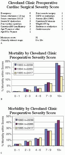
In looking at factors associated with a higher risk of death and postoperative morbidity, Higgins et al. developed and validated a severity scoring system using measurable factors that defined a patient population at increased risk (Table 24.6) (Figs. 24.2A-24.2C) (21). This Cleveland Clinic Preoperative Cardiac Surgical Severity Score assigned points (from 1 to 6), for the factors most closely associated with mortality and morbidity (21). As the severity score index became greater than 4, the incidence of morbidity and mortality markedly increased. The highest number of points was assigned to emergent surgery (6) while serum creatinine > 1.9 mg/dl was given 4 points. Severe LV dysfunction, prior cardiac surgery, and mitral valve insufficiency were assigned 3 points each. In correlating the total severity score with morbidity and mortality outcomes, patients with a total score ≥ 4, have a higher risk for postoperative morbidity and mortality when undergoing myocardial revascularization (21).


IMPORTANCE OF ISCHEMIC HEART DISEASE IN FUTURE HEALTH-CARE DELIVERY

Despite recent advances in the management of ischemic heart disease, the National Center for Health Statistics and World Health Organization report that it
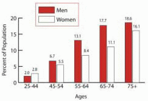
remains the leading cause of death throughout the world and will remain so through the year 2020 (22,23). The factors behind these trends are related to the increased incidence of coronary artery disease and associated risk factors in aging patients (for example, diabetes, hypertension, and obesity). The number of individuals over the age of 65 is expected to increase from 35 million to 71 million by the year 2030 in the United States alone (Fig. 24.3) (24). Of individuals over the age of 65, greater than 65% have cardiovascular disease (Fig. 24.4) (22,25). This age group has an increasing incidence of atherosclerotic vascular disease. More alarming is the increasing incidence of these factors in younger generations, suggesting that the cardiovascular epidemic will continue for years to come. Consequently, we find ourselves in the midst of a growing cardiovascular pandemic in the United States and throughout the world, with ischemic heart disease constituting the major cause of significant cardiovascular morbidity and mortality (26). In addition, 80% of the elderly over 60 years old have at least one chronic disease and 50% have two chronic diseases (23). In addition to these demographic trends, the number of percutaneous revascularizations will continue to increase. Patients with coronary artery disease are living longer. This has resulted in a surgical revascularization population that is older, has more diffuse coronary disease, more frequent complications associated with chronic ischemic disease, and a more frequent history of previous surgical interventions (PCI and CABG).


TABLE 24.6. Coronary Artery Bypass Surgery Cleveland Clinic Severity Score (21)


Age 65 to 74 years


1


Age 75 years or older


2


Weight ≤ 65 kg


1


Emergency


6


Severe LV dysfunction


3


Operative AV stenosis


1


Operative MV insufficiency


3


Prior cardiovascular surgery


2


Prior cardiac operation


3


Diabetes on medication


1


COPD on medication


2


Cerebrovascular


1


Serum creatinine 1.6-1.8 mg


1


Serum creatinine >1.9 mg


4


Anemia (Hct ≤ 34%)


2


Maximum score:


31


FIGURE 24.2. A. The Cleveland Clinic Severity Score was developed and validated in 1992 to predict those patients who were at risk for significant morbidity and mortality based on preoperative risk factors. B and C. Those patients with a severity score of 4 represent a patient population at higher risk of developing significant perioperative morbidity and increased mortality.


FIGURE 24.3. The aging of the United States population. It is estimated by the Center for Health Statistics that there will be more than 52 million individuals over the age of 65 living in the United States.


FIGURE 24.4. Age and incidence of coronary artery disease in men and women. The Center for Disease Control and Prevention reports that there is an age-related increase in the incidence of coronary artery disease.

The morbidity and mortality associated with myocardial revascularization has steadily declined over the last 35 years (28). If we are to maintain this trend, it will be necessary to develop strategies for addressing those critical issues that guide the intraoperative decision-making process. The purpose of this chapter is to provide an overview of the role of intraoperative echocardiography in the management of patients undergoing surgical revascularization of the myocardium. The principles of the intraoperative echo exam will be identified. The critical issues that must be addressed to insure the successful management of higher risk patients who are undergoing surgical myocardial revascularization will be examined, followed by outcome studies characterizing the effectiveness of such perioperative strategies. We will then focus on the complications of ischemic heart disease and their echocardiographic diagnosis and intraoperative management as guided by TEE. This discussion will conclude with an examination of future clinical applications of intraoperative TEE.


PRINCIPLES OF PERFORMING INTRAOPERATIVE ECHO EXAM IN CABG SURGERY

The intraoperative echo examination for higher risk patients undergoing myocardial revascularization is guided by principles related to the unique demands of the environment and the potential for sudden changes in the patient’s cardiovascular function (Table 24.7). Because of this potential for change in cardiovascular function throughout the procedure, complete diagnostic TEE exams are performed at each of the progressive phases of the surgical procedure:

1. Pre-CPB

2. Pre-Sep from CPB

3. Post-Sep CPB

4. Post-chest closure


TABLE 24.7. Principles of Intraoperative Echo Exam Higher Risk Coronary Artery Bypass Surgery


1.


Addresses critical issues


2.


Systematic examination


Organized by priority


Initial overview exam


Focused diagnostic exam


Comprehensive exam documented


3.


Efficient


4.


Severity assessment by weighted integration


5.


Study results recorded and discussed with surgeon


6.


Comprehensive digital study archived


7.


Results compared to preoperative data


Variances addressed


Communicated with surgical team


Potential Rx alterations discussed with cardiologist


8.


Qualified personnel trained and credentialed


9.


IOE exam under CQI process


10.


Equipment maintained and updated


In addition, should the patient encounter hemodynamic instability at any point during the course of the procedure, an overview is performed followed by a more “focused diagnostic exam” directed by the clinical course and overview exam. The examination following chest closure insures that grafts are not kinked, interrupting flow and impeding an accurate determination of the effect of chest closure on preload.

The intraoperative TEE exam is performed at each of the stages in a sequence that first resolves those critical issues guiding the patient’s management, yet, recognizes the ongoing management of the patient and the need to document a complete examination for future comparison. To prevent significant mid-exam revelations from occurring, an abbreviated 60-second overview exam may be performed followed by the remainder of the exam. In addition to the diagnostic issues that are already on the agenda, the abbreviated overview exam provides an up-to-date assessment of additional issues that might be addressed.

Because of the inherent difficulties in distinguishing degrees of degenerative calcification and fibrosis of the aorta and heart, subdued lighting in the OR reduces monitor glare, enabling a more accurate assessment of aortic arteriosclerosis, myocardial fibrosis, and calcification of cardiovascular structures. Periods without electrocautery interference during critical portions of the intraoperative exam also contribute to the acquisition of quality two-dimensional (2-D) images and Doppler-derived hemodynamic data. Such an atmosphere permits the precise recognition of intricate structural abnormalities (vegetation, thrombi, right-to-left shunts), which may have potentially devastating consequences if missed. It also enables the acquisition of the quality of diagnostic information, which may confidently guide the pivotal surgical and hemodynamic decisions.

The conclusions of the TEE exam are communicated directly to the surgical team in addition to being documented in the patient’s permanent medical record. Digital loops and images, which support the diagnostic conclusions and constitute the complete systematic examination, are achieved for future retrieval for comparison and reviewed under an organized CQI process.


Critical Issues in CABG Surgery Addressed by Intraoperative Echo (Tables 24.8A and 24.8B)

The purpose of the intraoperative echo is not to replace the patient’s preoperation assessment, but to confirm and refine it. Due to the clinical dynamic associated with ischemic heart disease, it is always possible that new ventricular or valve dysfunction may occur as a consequence of intervening ischemia or infarction. In addition to providing an ongoing method of monitoring the patients’ cardiovascular function, intraoperative echocardiography is used to address a number of critical issues that may influence the outcome of coronary bypass surgery. These
critical issues include the diagnosis of previously unrecognized cardiovascular abnormalities requiring additional unplanned surgical intervention or altering the patient’s anesthetic-hemodynamic management, development of the cannulation-perfusion strategy to prevent neurologic dysfunction, an assessment of the results of the surgical procedure and potential complications, and a final documentation of a comprehensive baseline study for future comparison. Each phase of the procedure has issues that are important to address depending on the extent of the patient’s preoperative assessment. If there is a concern regarding the viability of a specific region of myocardium, a pre-CPB dobutamine stress test may be performed as discussed in Chapter 23, Assessment of Myocardial Viability (29,30).


TABLE 24.8A. Critical Issues in Higher Risk Coronary Artery Bypass Surgery


1.


Intraoperative monitoring


2.


Diagnosis of unrecognized abnormalities changing management


Unplanned surgical procedures


Anesthetic-hemodynamic management


3.


Cannulation and perfusion strategy


4.


Predict post-CPB complications


5.


Surgical results and potential complications


Global and regional function


Functioning bypass grafts


Inotropic support


Mechanical support (IABP or LVAD / RVAD ECMO)


Valve function


Ischemic MR


TR


AR (LV distension prior to separation)


Complications of cannulation


Aortic dissection


Plaque disruption


Complications of myocardial protection


Regional dysfunction (septal)


RV dysfunction


Coronary sinus trauma


6.


Documentation of comprehensive study (future reference)


Global and regional (LV and RV) function


Valve function


LA, RA, and shunt potential


Aorta (ascending, arch, and descending)


TABLE 24.8B. Critical Surgical Decisions in Higher Risk Coronary Artery Bypass Surgery


Impact of Echo on Surgical Decision Making


Echo Finding


Surgical Decisions


Proximal aortic mobile atheroma


Off pump vs. on CPB


Alternative arterial cannulation (axillary, femoral)


Identify cross-clamp site (epiaortic echo)


Identify ascending aortic cannulation site


Identify site for antegrade cardioplegia insertion


Calcified atheromatous aorta


Off-pump CABG


All arterial grafts (IMA, gastroepiploic)


Axillary cannulation


Circulatory arrest


Significant aortic regurgitation


LV venting


Direct coronary administration of cardioplegia


Demonstrable regional viability


Coronary bypass graft of anatomy suitable


Pre-CPB


Additional unplanned bypass grafts


New regional wall motion abnormalities


Pre-CPB IABP, additional grafts, check angio


New valve dysfunction


Unplanned MVREP or MVR


Unplanned AVR, TVREP, or TVR


Post-CPB new regional wall motion abnormalities


Delayed wean from CPB, revision of grafts


IABP


Postbypass global LV dysfunction


Return to CPB


Additional grafts


Graft revision


IABP


LVAD


Post-CPB localized aortic dissection


Localized repair


Post-CPB extensive type I or II aortic dissection


Replace ascending aorta


Reduced celiac axis flow (metabolic acidosis)


Explore abdomen for gut ischemia


Mechanical complications of ischemic heart disease


VSD, aneurysm, pseudoaneurysm


Repair of structural defect


LV or LA thrombus


Thrombus removal


ischemic MR (ruptured papillary muscle)


MVR or MVRep


Intraoperative Monitoring

There are a number of determinants of cardiac function that may be monitored intraoperatively by TEE (Table 24.9A), including preload, diastolic function, contractility, regional myocardial function, valve function, and afterload. Compared with the hemodynamics and calculations derived from measurements obtained with the pulmonary artery thermodilution catheter, TEE provides a more direct physiologic assessment of each determinant of cardiac function (Table 24.9B). Hemodynamic instability is more often encountered in higher risk patients. It may be caused by hypovolemia, ventricular dysfunction, or low systemic vascular resistance. In these circumstances, the intraoperative echo provides a rapid method of assessing global (LV and RV) ventricular function, preload, presence of segmental dysfunction indicating myocardial ischemia, and an indication of lower systemic vascular resistance (low MAP with hypercontractile LV).

Two-dimensional echocardiography provides both qualitative and quantitative evaluation of systolic ventricular function. The midesophageal four-chamber (ME 4-chamber) and two-chamber (ME 2-chamber) views,
the midesophageal long-axis (ME LAX) view and transgastric short-axis (TG SAX) views (basal, midpapillary, and apical), and the transgastric long-axis view (TG LAX) allow assessment of global and regional ventricular function. Two-dimensional quantitative measures of LV systolic function include ventricular dimensions, volume, stroke volume (SV), cardiac output (CO), ejection fraction (EF), and regional wall motion abnormalities. Left ventricular ejection fraction (LVEF) can be calculated from LV end-systolic volume (LVESV) and end-diastolic volume (LVEDV) as the ratio (LVEDV-LVESV)/LVEDV. Cardiac output can be calculated using the area-length formula across a cardiac valve (most commonly the aortic valve): CO = 0.785 × LVOT D2 × LVOTVTI × HR (LVOT = left ventricular outflow tract; D = LVOT diameter; LVOTVTI = velocity time integral, measured with Doppler spectrum across the aortic valve; and HR = heart rate). IOE determination of CO using TEE is helpful even when a pulmonary artery catheter is used, because the thermodilution technique is inaccurate in patients with significant tricuspid valve regurgitation (31).


TABLE 24.9A. Intraoperative Assessment of Cardiac Function


Determinant


TEE


PA Catheter


Preload


Direct volume assessment


PAOP


PW Doppler pulmonary veins


RVEDP


Interatrial septum shift


CVP


Diastolic Function


Inflow patterns


Indirect PAOP


Tissue Doppler


Global Contractility


LV/RV 2-D image


Cardiac


output


Cardiac output by volumetric flow


Stroke volume


Regional Function


Systolic thickening


NA


Contraction pattern


Valve Function


2-D imaging of structure


cv wave


CF Doppler assessment


Afterload


Calculation regional wall stress


SVR calculation


Tissue Doppler imaging


Only gold members can continue reading. Log In or Register to continue

Stay updated, free articles. Join our Telegram channel

Jul 15, 2016 | Posted by in CARDIOLOGY | Comments Off on Assessment in Higher Risk Myocardial Revascularization and Complications of Ischemic Heart Disease

Full access? Get Clinical Tree

Get Clinical Tree app for offline access