Assessment


Assessment


Obtaining a health history

The first step in assessing the cardiovascular system is to obtain a health history. Begin by introducing yourself and explaining what will occur first during the health history and later during the physical examination. Then ask about the patient’s chief complaint. Also, be sure to ask about the patient’s personal and family health history.


Chief complaint

You’ll find that patients with cardiovascular problems typically cite specific complaints, such as chest pain, palpitations, syncope, intermittent claudication, and peripheral edema. Let’s take a closer look at each of these chief complaints as well as some other common signs and symptoms.


Chest pain

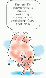
Many patients with cardiovascular problems complain at some point of chest pain. Chest pain can arise suddenly or gradually and can radiate to the arms, neck, jaw, or back. It can be steady or intermittent, mild or acute, and it can range in character from a sharp, shooting sensation to a feeling of heaviness, fullness, or even indigestion. Patients may deny pain but will describe chest discomfort which presents in diverse ways.


image


A full menu of causes

The cause of chest pain may be difficult to determine at first. Chest pain can be provoked or aggravated by stress, anxiety, exertion, deep breathing, or eating certain foods. (See Table 2.1, Understanding chest pain.)1


Table 2.1: Understanding chest pain


Use this table to help you more accurately assess chest pain and its possible causes.


What it feels like


Where it’s located


What makes it worse


What causes it


What makes it better


Aching, squeezing, pressure, heaviness, burning pain; usually subsides within 10 minutes


Substernal; may radiate to jaw, neck, arms, and back


Eating, physical effort, smoking, cold weather, stress, anger, hunger, supine position


Angina pectoris


Rest, nitroglycerin (Note: Unstable angina appears even at rest)


Tightness or pressure; burning, aching pain; possibly accompanied by shortness of breath, diaphoresis, weakness, anxiety, or nausea; sudden onset; lasts 30 minutes to 2 hours


Typically across chest but may radiate to jaw, neck, arms, and back


Exertion, anxiety


Acute coronary syndromes which include acute myocardial infarction and unstable angina2


Opioid analgesics such as morphine, nitroglycerin


Sharp and continuous pain; may be accompanied by friction rub; sudden onset


Substernal; may radiate to neck and left arm


Deep breathing, supine position


Pericarditis


Sitting up, leaning forward, anti-inflammatory drugs


Excruciating, tearing pain; may be accompanied by blood pressure difference between right and left arm; sudden onset


Retrosternal, upper abdominal, or epigastric; may radiate to back, neck, and shoulders


Not applicable


Dissecting aortic aneurysm


Opioid analgesics, surgery


Sudden, stabbing pain; may be accompanied by cyanosis, dyspnea, or cough with hemoptysis


Anterior and posterior thorax


Inspiration


Pulmonary embolus


Analgesics


Sudden and severe pain; sometimes accompanied by dyspnea, increased pulse rate, decreased breath sounds, or deviated trachea


Lateral thorax


Normal respiration


Pneumothorax


Analgesics, chest tube insertion


Dull, pressure-like, squeezing pain


Substernal, epigastric areas


Food, cold liquids, exercise


Esophageal spasm


Nitroglycerin, calcium channel blockers


Sharp, severe pain


Lower chest or upper abdomen


Eating a heavy meal, bending, supine position


Hiatal hernia


Antacids, walking, semi-Fowler’s position


Burning feeling after eating; sometimes accompanied by hematemesis or tarry stools; sudden onset that generally subsides within 15 to 20 minutes


Epigastric area


Lack of food, eating highly acidic foods


Peptic ulcer


Food, antacids


Gripping, sharp pain; may be accompanied by nausea and vomiting


Right epigastric or abdominal areas; may radiate to shoulders


Eating fatty foods, supine position


Cholecystitis


Rest and analgesics, surgery


Continuous or intermittent sharp pain; possibly tender to touch; gradual or sudden onset


Anywhere in chest


Movement, palpation


Chest wall syndrome/costochondritis


Time, analgesics, heat applications


Dull or stabbing pain; usually accompanied by hyperventilation or breathlessness; sudden onset; can last less than a minute or as long as several days


Anywhere in chest


Increased respiratory rate, stress, anxiety


Acute anxiety


Slowing of respiratory rate, stress relief


Pinpointing pain

If the patient’s chest pain isn’t severe, proceed with the history. Ask if the patient feels diffuse pain or can point to the painful area. Ask whether he has any discomfort that radiates to his neck, jaw, arms, or back. If he does, ask him to describe it. Is it a dull, aching, pressure-like sensation? A sharp, stabbing, knifelike pain? Does he feel it on the surface or deep inside? Ask him to rate the pain on a scale of 1 to 10, in which 1 means negligible and 10 means the worst imaginable.


Going steady?

Then find out whether the pain is constant or intermittent. If it’s intermittent, how long does it last? Ask if movement, exertion,
breathing, position changes, or eating certain foods worsens or helps relieve the pain. Does anything in particular seem to bring it on? Find out what medications the patient is taking, if any, and ask about recent dosage or schedule changes.


Palpitations

Defined as a conscious awareness of one’s heartbeat, palpitations are usually felt over the precordium or in the throat or neck. The patient may describe them as pounding, jumping, turning, fluttering, or flopping. He may also describe a sensation of missed or skipped beats. Palpitations may be regular or irregular, fast or slow, paroxysmal or sustained.


Don’t skip this beat

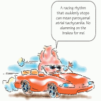
To help characterize the palpitations, ask the patient to simulate their rhythm by tapping his finger on a hard surface. An irregular “skipped beat” rhythm points to premature ventricular contractions, whereas an episodic racing rhythm that ends abruptly suggests paroxysmal atrial tachycardia (brief periods of tachycardia alternating with normal sinus rhythm).


image


Maybe it was that triple shot of espresso

Next, ask if the patient has a history of hypertension or if he has recently started digoxin therapy. Be sure to obtain a drug history, and ask about caffeine, tobacco, and alcohol consumption and use of illicit drugs or herbal supplements. Do not forget to ask about energy drinks. Palpitations may accompany use of these substances.


No big deal—unless…

Palpitations are typically insignificant and are relatively common. However, they can be caused by such cardiovascular disorders as arrhythmias, hypertension, mitral prolapse, and mitral stenosis.


Syncope

Syncope is a brief loss of consciousness caused by a lack of blood to the brain. It usually occurs abruptly and lasts for seconds to minutes. It may result from such cardiovascular disorders as aortic arch syndrome, aortic stenosis, and arrhythmias.


Barely breathing

When syncope occurs, the patient typically lies motionless, with his skeletal muscles relaxed. The depth of unconsciousness varies—some patients can hear voices or see blurred outlines; others are
unaware of their surroundings. The patient is strikingly pale with a slow, weak pulse; hypotension; and almost imperceptible breathing.


Fainting facts

If the patient reports a fainting episode, gather information about the episode from him and his family. Did he feel weak, lightheaded, nauseated, or diaphoretic just before he fainted? Did he get up quickly from a chair or from lying down? Any visual changes, such as blurring or narrow visual field? During the fainting episode, did he have muscle spasms or incontinence? How long was he unconscious? When he regained consciousness, was he alert or confused? Did he have a headache? Has he fainted before? If so, how often do the episodes occur?


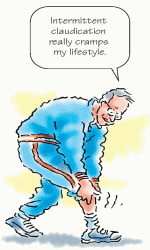
Intermittent claudication

Intermittent claudication is cramping limb pain that’s brought on by exercise and relieved by 1 or 2 minutes of rest. It most commonly occurs in the legs. This pain may be acute or chronic. When pain is acute and not relieved by rest, it may signal acute arterial occlusion.


Midlife crisis

Intermittent claudication is most common in men ages 50 to 60 who have a history of diabetes mellitus, hyperlipidemia, hypertension, or tobacco use. It typically results from such cardiovascular disorders as aortic arteriosclerotic occlusive disease, acute arterial occlusion, or arteriosclerosis obliterans.


Claudication interrogation

If the legs are affected, ask the patient how far he can walk before pain occurs and how long he must rest before it subsides. Can he walk as far as he could before, or does he need to rest longer? Does the pain-rest pattern vary? Is the pain in one leg or both? Where is the pain located? Has the pain affected his lifestyle?


Peripheral edema

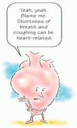
Peripheral edema results from excess interstitial fluid in the arms or legs. It may be unilateral or bilateral, slight or dramatic, pitting or nonpitting.


image


In your face (and arm and leg)

Arm and facial edema may be caused by superior vena cava syndrome or thrombophlebitis. Leg edema is an early sign of right-sided heart failure, especially if it’s bilateral. It can also signal thrombophlebitis and chronic venous insufficiency.


Since when?

Ask the patient how long he has had the edema. Did it develop suddenly or gradually? Does the edema decrease if the patient elevates his arms or legs? Is it worse in the mornings, or does it get progressively worse during the day? Did the patient recently injure the affected extremities or have surgery or an illness that may have immobilized him? Does he have a history of any cardiovascular disease? Is he taking medications? Which drugs has he taken in the past?


Personal and family health

After you’ve asked about the patient’s chief complaint, then inquire about his family history and past medical history, including heart disease, diabetes, and chronic lung, kidney, or liver disease. (See Recognizing cardio risk.)


All in the family

Ask if any family members have had heart disease, a history of myocardial infarction (MI), heart failure, cerebrovascular accident (CVA), or an unexplained, sudden death. Find out at what age the MIs occurred.


Getting personal

In addition to obtaining information about the patient’s family history, be sure to ask the patient about his:

• stress level and coping mechanisms

• current health habits, such as smoking and exercise habits, alcohol and caffeine intake, and dietary intake of fat and sodium

• drug use, including over-the-counter drugs, illicit drugs, and herbal supplements


• previous operations

• environmental or occupational hazards

• activities of daily living.


Also related

Also ask the patient these questions:

• Are you ever short of breath? If so, what activities cause you to be short of breath?

• How many pillows do you use for sleep?

• Do you feel dizzy or fatigued?

• Do your rings or shoes feel tight?

• Do your ankles swell?

• Have you noticed changes in color or sensation in your legs?

If so, what are those changes?

• If you have sores or ulcers, how quickly do they heal?

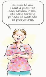
• Do you stand or sit in one place for long periods at work?


image


Performing a physical assessment

The key to accurate assessment is regular practice, which helps improve technique and efficiency. A consistent, methodical approach to your assessment can help you identify abnormalities.


Shopping list

For the physical assessment, you’ll need a stethoscope with a bell and a diaphragm, an appropriate-sized blood pressure cuff, a ruler,
and a penlight or other flexible light source. Make sure the room is quiet.


Dressed down

Ask the patient to remove all clothing except his underwear and to put on an examination gown. Have the patient lie on his back, with the head of the examination table at a 30- to 45-degree angle. Stand on the patient’s right side for the exam since you may need the patient to assume a left lateral recumbent position to move the heart closer to the chest wall.3


Assessing the heart

During your assessment, inspect, palpate, percuss, and auscultate the heart.


Inspection

First, take a moment to assess the patient’s general appearance. Is he overly thin? Obese? Alert? Anxious? Note his skin color, temperature, turgor, and texture. Are his fingers clubbed? If the patient is dark-skinned, inspect his mucous membranes for pallor.


Checking out the chest

Next, inspect the chest. Note landmarks you can use to describe your findings and to identify structures underlying the chest wall. (See Identifying cardiovascular landmarks.) Also, look for pulsations, symmetry of movement, retractions, or heaves (a strong outward thrust of the chest wall that occurs during systole).


image


Location, location

Position a light source, such as a flashlight or gooseneck lamp, so that it casts a shadow on the patient’s chest. Note the location of the apical impulse. You should find it in the fifth intercostal space, medial to the left midclavicular line. Because it corresponds to the apex of the heart, the apical pulse helps indicate how well the left ventricle is working. The apical pulse is usually the point of maximal impulse (PMI). Remember, though, that the apical impulse can be seen only in about 50% of adults. You’ll notice it more easily in children and in patients with thin chest walls. To find the apical impulse in a woman with large breasts, displace the breasts during the examination.

Jun 7, 2016 | Posted by in CARDIOLOGY | Comments Off on Assessment

Full access? Get Clinical Tree

Get Clinical Tree app for offline access