Approach to and Management of Chronic Venous Insufficiency


Approach to and Management of Chronic Venous Insufficiency


Justin B. Hurie

Thomas W. Wakefield


Chronic venous insufficiency (CVI) is a pathologic condition of lowerextremity venous hypertension often due to dysfunction of the vein walls or valves. The most common etiologies of vein dysfunction are primary valvular incompetence and secondary dysfunction due to a prior deep vein thrombosis (DVT), with DVT occurring in more than 350,000 patients annually. The incidence of CVI after appropriate anticoagulation may be as high as 23% after 2 years and 28% after 5 years. Less common causes include cavernous hemangiomas, congenital arteriovenous fistulas, and pelvic tumors. Varicose veins are the most common manifestation of CVI, affecting an estimated 20 million people. Other symptoms include edema, hyperpigmentation, lipodermatosclerosis, and ulceration. The overall incidence of venous stasis syndrome is 76 per 100,000 personyears and rises with increasing age. Although advanced CVI symptoms such as venous ulcers are much less common, having an incidence of 18 per 100,000 person-years, the economic impact is nonetheless significant with an estimated $200 million to $1 billion per year being spent on their treatment in the United States. The prevention, diagnosis, and treatment of this disease process, therefore, become highly relevant.


CLINICAL FEATURES


History and Physical Examination

Symptoms of CVI are variable and include leg tiredness, cramping, itching, burning, and swelling. Physical findings may include prominent dilated superficial veins, edema, skin changes, and ulceration.


Presenting Features

The typical presentation of a patient with CVI is one of edematous lower extremities (unilateral or bilateral) with hyperpigmentation, eczema, and/or lipodermatosclerosis (scarring of the skin and fat). Accompanying varicose veins or skin ulceration may also be present. Edema is the initial finding and most commonly occurs at and above the ankle, without involvement of the forefoot or toes. It usually responds to leg elevation and is typically worse in the evening. When ulcers occur, they are most often large, irregular, and associated with a shallow moist granulation base (Fig. 22.1). They are often very painful and occur on the medial or lateral surface of the leg in the supramalleolar position. Because of the significant variability in the manifestations and severity of CVI, several classification schemes have been designed to help categorize patients. Two such schemes will be briefly reviewed later in this chapter.


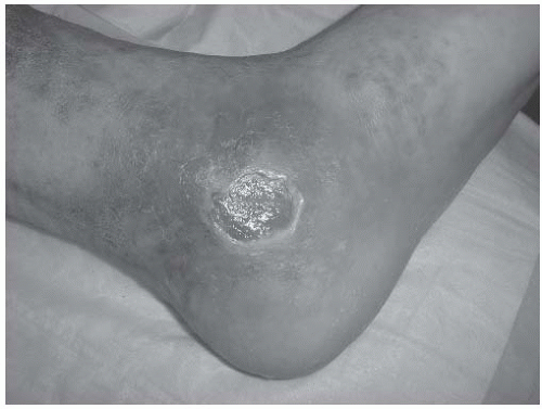
FIGURE 22-1. Chronic venous stasis ulcer with location in the medial paramalleolar position.


Differential Diagnosis

The symptoms of CVI (pain, edema, skin changes, and ulcerations) must be distinguished from other etiologies of these symptoms. Likewise, venous ulcers can be mistaken for other lesions such as squamous cell carcinoma (Marjolin’s ulcers).


Pathophysiology

The pathophysiology of CVI can be broken down into changes occurring at the level of the large venous vessels, the microvasculature, and the tissue. CVI occurs as a result of valvular dysfunction within the large veins of the leg. The resulting standing column of blood leads to venous hypertension. Venous hypertension is defined as end-exercise venous pressure at the ankle greater than 30 mm Hg (normal is 15 mm Hg). This, in turn, leads to capillary dysfunction and tissue injury. Common causes of valvular dysfunction include primary valvular incompetence and secondary valvular incompetence after DVT. In patients with no history or evidence of DVT, primary valve abnormalities such as valvular agenesis or aplasia are most likely, although vein wall abnormalities can also lead to valvular insufficiency. Contemporary data in DVT patients suggest that the incidence of severe CVI after 8 years follow-up is approximately 30% and may be even higher if associated with ipsilateral recurrent DVT. It has also been shown that valvular dysfunction occurs more commonly if there is delayed DVT resolution and if the more proximal veins are involved in the initial venous thrombosis. Skin ulceration is also more common in patients with recurrent DVT. Risk factors for DVT formation can be inherited, acquired, or multifactorial (Table 22.1). Once valvular dysfunction occurs, the ensuing chronic venous hypertension has profound effects at the microvascular level. Capillaries become dilated and
tortuous, allowing extravasation of fluid, red blood cells, and macromolecules. There is an increase in white blood cell adhesion to the endothelium as well as leukocyte activation. Increased microvascular blood flow is seen initially, but there can be late findings of capillary thrombosis leading to tissue hypoxia. Within the tissues, the extravasated red blood cells break down to produce a dark pigment called hemosiderin. The perivascular space becomes surrounded by intracellular matrix proteins. Chronic accompanying inflammation leads to scarring and fibrosis of the subcutaneous tissues. Macrophages, mast cells, and T lymphocytes are the cell types most commonly involved in tissue destruction. As skin perfusion decreases, the resulting ischemia can lead to skin breakdown and ulceration.


TABLE 22.1 RISK FACTORS FOR DVT


Congenital


Acquired


Situational


Mixed


Factor VII excess


HIV infection


Surgery


Hyperhomocysteinemia


Protein C deficiency


Age


Trauma


Increased factor VIII levels


Protein S deficiency


Immobilization


Pregnancy


Increased factor IX levels


Factor V Leiden


Malignancy


Oral contraceptives


Increased factor XI levels


Antithrombin deficiency


Heparin-induced thrombocytopenia


Hormone replacement therapy


Prothrombin G20210A


Behcet’s disease


Nephrotic syndrome


Antiphospholipid antibodies


Myeloproliferative disorders


Polycythemia vera


Paroxysmal nocturnal hemoglobinuria


Inflammatory bowel disease


(From Gloviczki P, ed. Handbook of venous disorders. London, UK: Arnold, 2009:95, with permission.)


Classification

As illustrated in the previous section, there are many manifestations of venous hypertension. Due to the variety of presentations, it is important to standardize the classification of degree of CVI. The most commonly used classification is the CEAP system illustrated in Table 22.2. In addition, the Villalta

scoring system and Venous Clinical Severity Score (VCSS) system have been described in order to objectively describe the clinical response. The Villalta scale gives 0 (none) to 3 (severe) points for five patient-given symptoms and six physician-observed signs. A score greater than 5 is consistent with the postthrombotic syndrome and has been shown to correlate with quality of life. The VCSS also awards 0 (none) to 3 (severe) for ten different complaints (see Table 22.3). These systems are recommended by various organizations including the American Venous Forum in order to standardize reporting and allow more objective observation of clinical benefit and improved research.


TABLE 22.2 CEAP CLASSIFICATION


Clinical Classification


CO


No visible or palpable signs of venous disease


C1


Telangiectasias or reticular veins


C2


Varicose veins


C3


Edema


C4a


Pigmentation and/or eczema


C4


Lipodermatosclerosis and/or atrophie blanche


C5


Healed venous ulceration


C6


Active venous ulceration


S


Symptoms including pain, tightness, skin irritation, heaviness, or muscle cramps


A


Asymptomatic


Etiologic Classification


Ec


Congenital


Ep


Primary


ES


Secondary (postthrombotic)


En


No venous etiology identified


Anatomic Classification


As


Superficial veins


1


Telangiectasias/reticular veins


2


Great saphenous vein (above knee)


3


Great saphenous vein (below knee)


4


Small saphenous vein


5


Nonsaphenous veins


Ad


Deep veins


6


Inferior vena cava


7


Common iliac vein


8


Internal iliac vein


9


External iliac vein


10


Pelvic veins (gonadal, broad ligament, other)


11


Common femoral vein


12


Deep femoral vein


13


Femoral vein


14


Popliteal vein


15


Crural vein (anteriortibial, posterior tibial, peroneal)


16


Muscular vein (gastrocnemial, soleal, other)


Ap


Perforating veins


17


Thigh perforator veins


18


Calf perforator veins


Pathophysiological


Classification


P1


Reflux


Po


Obstruction


Pr,o


Reflux and obstruction


Pn


No venous pathophysiology identified


(From Gloviczki P, ed. Handbook of venous disorders. London, UK: Arnold, 2009:38-39, with permission.)


DIAGNOSIS


Diagnostic Modalities

Several diagnostic studies may be employed in the evaluation of CVI, and they can be divided into invasive and noninvasive tests. A careful history and physical examination including arterial evaluation form the cornerstone of diagnostic evaluation. Within the last 10 years, duplex ultrasound has become the primary noninvasive diagnostic modality due to high sensitivity and specificity rates greater than 95%. Duplex ultrasound is safe during pregnancy and may also elucidate other causes of the patients’ symptoms. Additional noninvasive tests may be performed selectively and include air plethysmography (APG). Magnetic resonance venography or invasive testing such as phlebography is usually reserved for the evaluation of proximal venous structures in patients in whom surgical treatment is being considered. Historically, the Brodie-Trendelenburg test has been performed as an in-office evaluation for patients with prominent varicose veins. This tourniquet-based test, which helps discriminate saphenofemoral junction (SFJ) incompetence from deep-to-superficial communication via incompetent perforators, has been supplanted by the use of ultrasoundbased testing and is not usually performed in modern practice. The duplex evaluation helps localize specific perforators that may be contributing to the varicosities along with other points of reflux and may discriminate obstruction from valvular incompetence and reflux.


Duplex Ultrasound

Duplex ultrasound scanning is the modality of choice for the diagnosis of both acute and chronic DVT as well as to evaluate the presence of reflux. Compression maneuvers and examination of flow patterns with augmentation can allow systematic evaluation of the saphenofemoral and saphenopopliteal junctions and deep, superficial, and perforating veins. Venous reflux is identified by having the patient do Valsalva maneuver while in the 15-degree reverse Trendelenburg position or by using rapidly deflating pneumatic cuffs below the level being evaluated to elicit reflux. Some recommend that this technique be performed in the upright position. Incompetent perforator veins are identified and their size is measured by holding the transducer directly over the vein while squeezing and releasing the leg. Bidirectionality of flow with compression and release indicates an incompetent perforating vein. Limitations of duplex ultrasound scanning

include the need for an experienced vascular technologist to perform the exam, interoperator variability, and the inability to accurately visualize the venous system above the inguinal ligament. Duplex ultrasound provides direct evaluation of the veins in the lower extremity and indirect evaluation of the pelvic vasculature. The evaluation of the iliac veins through ultrasound relies on indirect evidence of obstruction such as loss of characteristic cessation of venous flow with Valsalva maneuver and loss of respiratory variation, indicating a more proximal obstruction.


TABLE 22.3 VENOUS CLINICAL SEVERITY SCORE


Attribute


Absent = 0


Mild = 1


Moderate = 2


Severe = 3


Pain


None


Occasional, not restricting activity or requiring analgesics


Daily, moderate activity limitation, occasional analgesics


Daily, severe limiting activities or requiring regular use of analgesics


Varicose veinsa


None


Few, scattered; branch veins


Multiple; GS veins confined to calf or thigh


Extensive; thigh and calf or GS and SS distribution


Venous edemab


None


Evening ankle edema only


Afternoon edema, above ankle


Morning edema above ankle and requiring activity change, elevation


Skin pigmentationc


None or focal, low intensity (tan)


Diffuse, but limited in area and old (brown)


Diffuse over most of gaiter distribution (lower 1/3) or recent pigmentation (purple)


Wider distribution (above lower 1/3) and recent pigmentation


Inflammation


None


Mild cellulitis, limited to marginal area around ulcer


Moderate cellulitis, involves most of gaiter area (lower 1/3)


Severe cellulitis (lower 1/3 and above) or significant venous eczema


Induration


None


Focal, circummalleolar (<5 cm)


Medial or lateral, less than lower third of leg


Entire lower third of leg or more


No. of active ulcers


0


1


2


>2


Active ulceration, duration


None


<3 mo


>3 mo, <1 y


Not healed > 1 y


Active ulcer, sized


None


<2-cm diameter


2-6-cm diameter


>6-cm diameter


Compressive therapy


Not used or not compliant


Intermittent use of stockings


Wears elastic stocking most days


Full compliance: stockings + elevation


a “Varicose” veins must be >4-mm diameter to qualify so that differentiation is ensured between C1 and C2 venous pathology.

b Presumes venous origin by characteristics [e.g., Brawny (not pitting or spongy) edema], with significant effect of standing/limb elevation and/or other clinical evidence of venous etiology (i.e., varicose veins, history of DVT). Edema must be a regular finding (e.g., daily occurrence).

c Focal pigmentation over varicose veins does not qualify.

d Largest dimension/diameter of largest ulcer.


GS, great saphenous; SS, small saphenous.


(Reproduced from Rutherford RB, Padberg FT, Comerota AJ, et al. Venous severity scoring: an adjunct to venous outcome assessment. J Vase Surg 2000;31:1307-1312, with permission.)


Air Plethysmography

An additional diagnostic study is APG, which is a physiologic study to measure venous reflux, calf muscle pump function, and venous obstruction. APG testing makes precise volume measurements with air-filled polyurethane sleeves that surround the legs. The first of several measurements that can be made with APG is the functional venous volume (VV). In normal limbs, the average VV is 80 to 150 mL, while in patients with severe CVI, the volume might be close to 400 mL. In order to measure VV, the patient is asked to stand from a supine position, and the VV is measured within the context of the venous filling index (VFI). The VFI is defined as the ratio between 90% VV and the time taken for 90% filling (VFT90) to occur (VFI = 90%VV/VFT90). In the normal situation, veins should fill slowly from the arterial side with a Vfiless than 2 mL/s. In limbs with severe reflux, this number may increase up to 30 mL/s. VFI has been shown to correlate with the prevalence of skin changes and ulceration. Calf muscle pump function is also assessed by APG. The patient is asked to do tiptoe movements, and the recorded decrease in pressure represents the ejected volume (EV). Thus, the ejection fraction (EF) can be measured with the formula EF = [(EV/VV) × 100]. A normal EF is above 60%. Severe deep venous disease can result in an EF below 10%. The EF, like the VFI, is an important predictor of which patients will develop skin ulceration. Finally, APG can also be used to measure obstruction using a thigh tourniquet. The benefit of APG in clinical practice is that it gives the physician an understanding of the different components contributing to the overall severity of patients’ CVI. APG can also be an important part of patient selection and predicting who will benefit most from surgical therapy. However, the most reliable part of APG is the assessment of reflux.


Phlebography

Ascending phlebography, once considered the “gold standard” in the assessment of chronic venous obstruction, is infrequently used given the advances in noninvasive testing. Phlebography is performed by injecting contrast into a dorsal foot vein and directing the contrast into the deep venous system by using an ankle tourniquet. This technique can provide useful anatomic information about the location and extent of venous obstruction. However, the profunda femoris veins and internal iliac veins are not typically well visualized with this technique.

An invasive technique used to assess venous reflux and valvular anatomy is descending phlebography. This test is performed on a fluoroscopic table
tilted 60 degrees, while contrast is injected through a catheter with the tip placed at the distal external iliac vein. Gravity and a Valsalva maneuver propel the contrast distally in limbs with incompetent valves. This allows grading of Phlebographie reflux as well as delineating competent valve cusps, which are clearly outlined with this technique. Reflux grades include 0, no reflux; 1, reflux into the femoral vein to the level of the proximal thigh; 2, reflux into the femoral vein but not through the popliteal vein; 3, reflux to a level just below the knee; and 4, reflux through to the level of the calf or ankle. However, descending phlebography requires an experienced imaging team to perform and is costly. Duplex scanning obviates the need for this test in many circumstances, although phlebograms are often performed in conjunction with therapeutic maneuvers such as venoplasty and stenting as well as thrombolysis.

Only gold members can continue reading. Log In or Register to continue

Stay updated, free articles. Join our Telegram channel

Jun 12, 2016 | Posted by in CARDIOLOGY | Comments Off on Approach to and Management of Chronic Venous Insufficiency

Full access? Get Clinical Tree

Get Clinical Tree app for offline access