Fig. 5.1
Standard approach for CABG
5.2 Mini Sternotomy
5.2.1 Mini Left Thoracotomy
With the spread of OPCAB, mini left anterior thoracotomy and OPCAB of the left anterior descending branch have come to be called the “left anterior small thoracotomy” (LAST) approach, as coined by Calafiore et al. [1]. When OPCAB is performed by a small incision method, it is defined as minimally invasive direct coronary artery bypass (MIDCAB) surgery, and MIDCAB and LAST are considered to be synonymous. CABG [2] is basically only performed by this method for diagonal branches including LAD, and when detachment of ITA is difficult, or the target vessel cannot be identified, the procedure has to be converted to full sternotomy; therefore, more institutes are performing full sternotomy from the start.
The body is placed at approximately 30° in a right lateral decubitus position. It is necessary to take a chest X-ray and a CT before the procedure to decide on the optimal intercostal space, but the skin incision is usually approximately 8 cm long at the fourth or fifth intercostal space (Fig. 5.2). The anterior chest muscle layer is incised, the pleura is opened from the outside, and once the lung has collapsed, the incision proceeds toward the midline, and the fourth or fifth costal cartilage is dissected for approximately 2 cm (Fig. 5.3). A small rib retractor is positioned, and while the intercostal space is slightly opened, the position of the LITA is confirmed (Fig. 5.4). If the rib retractor is opened widely at this time, care should be taken not to damage LITA by pulling it. While carefully cauterizing small branches with electrocautery, the rib retractor is gradually opened to free the LITA from the cranial and the caudal side over the width of one rib. Adequate working space can then be assured by positioning a special rib retractor or traction hook (Fig. 5.5). After the LITA has been detached, the pericardium is incised, and the target coronary artery is then identified (Fig. 5.6).
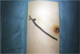
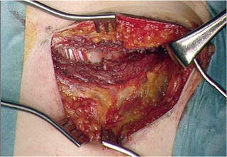
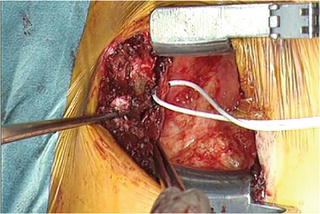
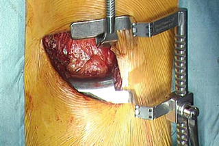
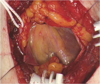

Fig. 5.2
An approximate 8 cm skin incision is made in the fourth or fifth intercostal space

Fig. 5.3
The anterior muscle layer of the chest is incised, and the pleura is opened from the outside

Fig. 5.4
A small rib retractor is inserted, and the position of LITA is confirmed while opening it slightly

Fig. 5.5
A special rib retractor or traction hook is used to secure adequate working space

Fig. 5.6
The pericardium is incised, and the target coronary artery is identified
5.2.2 Lower-End Sternal Splitting Approach
In order to overcome the problem of detaching the ITA and exposing the target vessel with mini left thoracotomy, Niinami et al. [3] have reported the lower-end sternal splitting (LESS) method using a small incision of the lower part that does not transverse the sternum. The body is positioned as with the standard median sternotomy method, and an approximate 8 cm skin incision from close to the 4th rib to a point slightly cranial of the xiphoid process is made. If the right gastroepiploic artery (RGEA) is used as the graft, the incision has to be extended caudally. The soft tissues close to the third intercostal space and underneath the xiphoid process are detached, and the lower part of the sternum is cut in the midline with an oscillating saw close to the third intercostal space. However, transverse incisions like a T-shaped or inverted L-shaped incision are not made in the sternum. By lifting the sternum with the retractor used to detach the ITA, it is possible to detach the ITA from close to the 2nd rib to close to the xiphoid process. When the LITA has been detached, the LAST rib retractor is used. Even without splitting the sternum transversely, the sternum has become pliable by lifting it to detach the LITA, and by opening the rib retractor gradually, the field of view can be expanded. Usually an adequate field of view from the root of the ascending aorta and the main pulmonary artery on the cranial side to the diaphragm on the caudal side can be obtained. Even if the ITA retractor is used to lift the sternum in this way, the ITA is not damaged, and the postoperative complication of a false sternal joint does not occur, and this method has the further advantage that the longitudinal incision of the sternum can simply be extended if it suddenly becomes necessary to change the procedure to a full sternotomy.
5.2.3 T-Shaped Incision
By the lower thoracic incision method [4], a skin incision is made from the third intercostal space to approximately 5 cm cranial from the xiphoid process, and a transverse incision is made at the height of the second intercostal space. By this method, with a longitudinal incision of approximately 10–12 cm of the sternum and a transverse incision at the height of the second intercostal space, it is possible to expose the right coronary artery (RCA) and LAD. Since the ascending aorta and right auricle can be visualized directly, it is possible to perform cannulation in this operative field and immediately start assisted circulation in case the hemodynamics become unstable.
Stay updated, free articles. Join our Telegram channel

Full access? Get Clinical Tree


