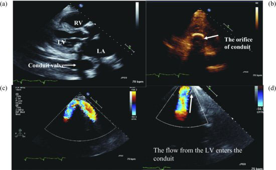
Figure 94.2 Parasternal long-axis view reveals a conduit below left ventricular posterior wall entering descending aorta (a). PAS view shows the orifice of conduit in apex (b). Apical view shows the conduit from the apex along left ventricular lateral wall (c). Apical view shows the flow entering conduit (d). LA, left atrium; LV, left ventricle; RV, right ventricle.
Magnetic resonance imaging reveals the entire apicoaortic conduit from LV apex entering descending aorta (Videoclip 94.2).
Discussion
In 1977, Reder et al.
Stay updated, free articles. Join our Telegram channel

Full access? Get Clinical Tree


