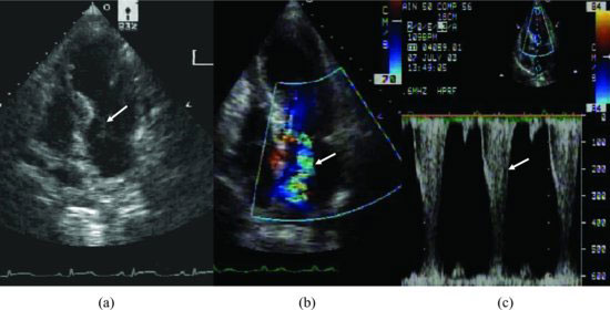
Figure 85.2 Apical 4-chamber view and continuous wave Doppler echocardiograms revealing extensive anteroapical akinesis and the basal function preserved, which lead to mitral anterior leaflet anterior movement during systole (a, arrow) and mitral regurgitation (b, arrow). The sequence of our patient’s presentation suggests that the apical ballooning caused geometric alterations of LV that in turn led to acute mitral regurgitation, systolic anterior motion, and mid left ventricular obstruction (c, high velocity).
Coronary angiography demonstrated nonobstructive coronary disease.
Discussion
Stay updated, free articles. Join our Telegram channel

Full access? Get Clinical Tree


