Figure 44.2 Aortopulmonary window (28 mm) can be well seen between the ascending aorta and the main pulmonary artery from the suprasternal fossa view (a). Color Doppler echocardiography shows the shunt from ascending aorta into pulmonary artery (b). AAO, ascending aorta; DAO, descending aorta; PA, pulmonary artery.
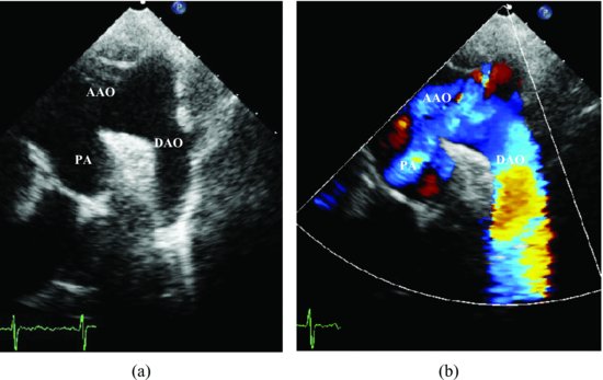
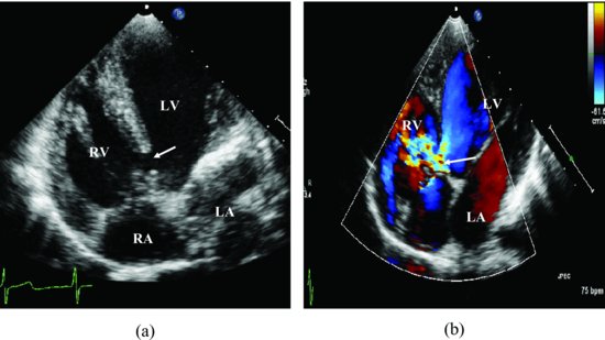
Figure 44.3 A perimembrane ventricular septal defect (arrow) detected from apical 4-chamber view (a). There was left-to-right shunt (arrow) through perimembrane ventricular septal defect during early systole (b). LA, left atrium; LV, left ventricle; RA, right atrium; RV, right ventricle.
Stay updated, free articles. Join our Telegram channel

Full access? Get Clinical Tree


